Figures & data
Table 1 Clinical manifestations seen with Leigh syndrome patients
Table 2 Genes known to be associated with Leigh syndrome
Figure 1 Axial MRI scan (A) and MRS spectrum (B) of a 9-year old boy with Leigh syndrome due to a mutation in the mtDNA.
Notes: (A) Axial fluid-attenuated inversion recovery image from a 3 T scanner (Siemens Trio) of a 9-year-old boy with Leigh syndrome resulting from a mitochondrial DNA mutation (m.11487 C>T). There is T2/fluid-attenuated inversion recovery hyperintensity in the bilateral caudate and putamen. Ex vacuo dilation of the frontal horns and bodies of the lateral ventricles is well visualized. (B) Summed spectrum from central gray nuclei in the same patient using the point-resolved spectroscopy pulse sequence (3 T Siemens Trio; time to echo (TE), 288 ms; time to relaxation (TR), 1,700 ms; 16×16 acquisition, interpolated to 32×32, 16 cm). The region of interest is the outlined region in the right putamen. An elevated lactate doublet peak (the lactate peak is upward at this TE time) is found at the echo time of 135 ms. N-acetylaspartate (NAA), choline-containing compounds (Cho), creatine + phosphocreatine (Cr), and second creatine (Cr2) peaks are also shown.
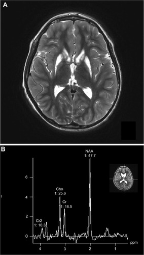
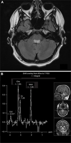
Figure 2 Axial MRI scan (A) and MRS spectrum (B) of a 17-year old male with Leigh syndrome due to a mutation in the mtDNA.
Notes: (A) Axial fluid-attenuated inversion recovery image from a 3 T scanner (Siemens Trio) of a 17-year-old teenage boy with Leigh syndrome resulting from a mitochondrial DNA mutation (m.3700 G>A). There is a T2/fluid-attenuated inversion recovery hyperintensity in the caudal pons. Not shown is the hyperintensity that extends to the cervicomedullary junction. There were no abnormalities noted in the basal ganglia. (B) Summed spectrum from the caudal pons in the same patient using the point-resolved spectroscopy pulse sequence (3 T Siemens Trio; TE, 135 ms; TR, 5,180; 16×16 acquisition, interpolated to 32×32 cm). The region of interest is the outlined region of the caudal pons. An elevated lactate doublet peak is found at the echo time of 135 ms (peak is downward at this TE time). There is a reduced N-acetylaspartate (NAA) peak suggesting loss of neuronal integrity in the same region. Choline-containing compounds (Cho), creatine + phosphocreatine (Cr), and second creatine (Cr2) peaks are also shown.