Figures & data
Figure 1 Schematic depiction of antisense-mediated splicing modulation approaches.
Notes: (A) Restoration of cryptic splicing for β-globin mutations. Due to a mutation in intron 3, a cryptic exon is included into the β-globin mRNA, abrogating the production of β-globin proteins. ASOs targeting the cryptic splice sites prevent the inclusion of the cryptic exon, restoring normal splicing and production of β-globin protein. (B) Restoration of cryptic splicing for USH1C. Due to a mutation in exon 3, a cryptic splice site within exon 3 is activated, leading to the exclusion of the last part of the exon from the USCH1C transcript and loss of harmonin protein production. Using ASOs targeting the cryptic splice site in exon 3 reactivates the original splice site and restores normal splicing and protein production. (C) Shifting of alternative splicing. The STAT3 gene produces STAT3α and STAT3β proteins based on the use of a proximal or more distal acceptor splice site in exon 23 during pre-mRNA splicing. ASOs targeting the proximal acceptor splice site lead to a shift into the production of the proapoptotic STAT3β protein. (D) Isoform-specific knockdown. The APOB gene gives rise to APOB100 and APOB48 proteins. The latter derives from RNA editing in exon 26. Skipping of exon 27 gives rise to a prematurely truncated APOB protein with cholesterol-lowering properties, without affecting the APOB48 isoform.
Abbreviations: APOB, apolipoprotein B100; ASOs, antisense oligonucleotides; LDL, low-density lipoprotein; mRNA, messenger RNA; STAT3, signal transducer and activator of transcription 3; USH1C, Usher syndrome type 1C gene.
Abbreviations: APOB, apolipoprotein B100; ASOs, antisense oligonucleotides; LDL, low-density lipoprotein; mRNA, messenger RNA; STAT3, signal transducer and activator of transcription 3; USH1C, Usher syndrome type 1C gene.
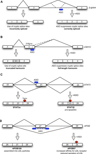
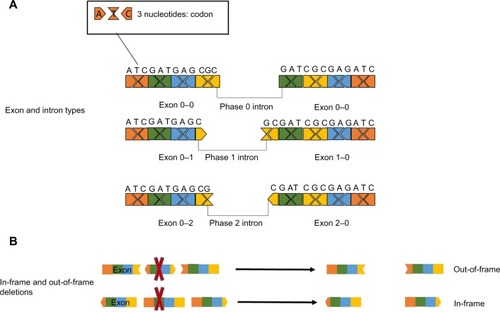
Figure 2 Effect of exon deletions on the open reading frame.
Notes: (A) Different ways in which codons in exons may be split by introns: in phase 0 introns, the codon ends at the end of the exon, while in phase 1 and phase 2 introns the codon is split over the 2 exons, with the interruption after the first or the second nucleotide of the codon, respectively. (B) As each exon is separated from the next and previous one by the flanking introns, there are nine different types of exons due to the different combinations (0–0, 0–1, 0–2, 1–0, 1–1, 1–2, 2–0, 2–1, 2–2); if one of these exons is deleted, the open reading frame could be either disrupted or maintained, depending on these combinations: the deletion of “symmetrical” exons (0–0, 1–1, 2–2) would have no effect on the reading frame (in-frame), while others will disrupt it (out-of-frame).