Figures & data
Figure 1 Positive lupus band test at low magnification: immunoglobulin class M deposits at the dermoepidermal junction in sun-protected nonlesional skin in a 26-year-old woman with systemic lupus erythematosus (original magnification: ×100).
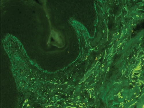
Figure 2 A sharply defined thin linear band at the dermoepidermal junction in pemphigoid (immunoglobulin class G deposits, original magnification:×200).
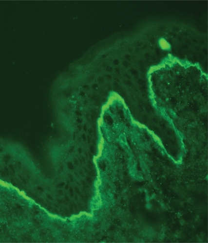
Figure 3 Partially homogenous, partially granular pattern of the lupus band test (immunoglobulin class M deposits, original magnification×200).
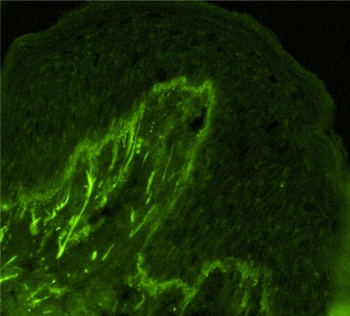
Figure 4 Stippled pattern of complement component 3 deposits in sun-protected nonlesional skin in patient with systemic lupus erythematosus (original magnification: ×400).
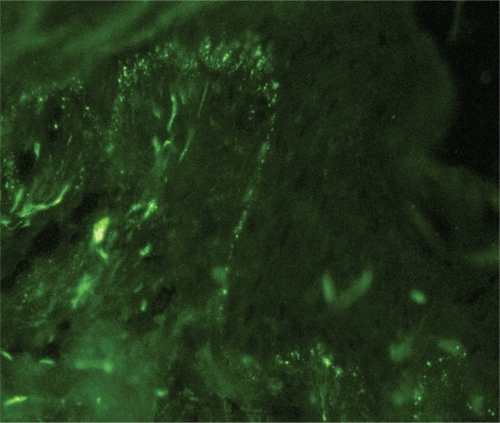
Figure 5 Shaggy pattern of complement component 3 deposits in sun-protected lesional skin in patients with subacute cutaneous lupus erythematosus.
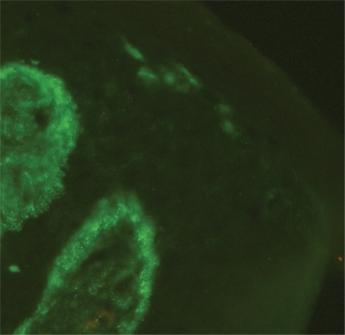
Figure 6 Granular (lumpy) pattern of complement component 3 deposits in sun-protected nonlesional skin in patient with systemic lupus erythematosus (original magnification:×400).
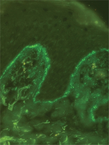
Table 1 Differential diagnosis of lupus band test in lupus erythematosus from other conditions