
Figures & data
Figure 1 A 17-year-old male with pain in the right upper abdomen.
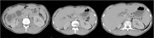
Figure 2 A 20-year-old male with pain in the right upper abdomen.
Abbreviation: CT, computed tomography.
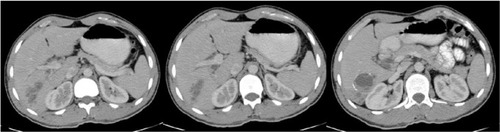
Table 1 The distribution of the cysts in the liver according to the Couinaud segmental anatomy and the relationship with CBC
Table 2 The distribution of the cysts in the liver according to lobe anatomy and the relationship with CBC
Table 3 The distribution of the cysts in the liver according to distance from the hilus and the ratio of CBC
Table 4 The distribution of the cysts according to the Gharbi classification and the ratio of CBC
Table 5 The relation of the demographic findings of the cysts and the patients and preoperative laboratory results in terms of CBC
Table 6 The linear regression analyses of predictive factors in determining CBC preoperatively