
Figures & data
Figure 1 Thorax CT manifestations of organizing pneumonia cases. Consolidation (A), mass like lesion (B), ground glass opacity (C), cavity (D).
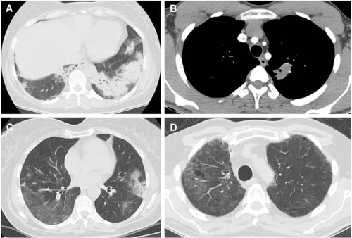
Figure 2 A foreign body tissue reaction against germinative membrane of probable hydatid cyst on histological examination of an excisional biopsy.
Notes: (A) There is a dense inflammatory reaction composed of lymphocytes, plasma cells, and histiocytes in the lung parenchyma surrounding the lesion (100× H&E). (B) In another area of the same slide, there are Masson bodies filling the airspaces (100× H&E). (C) Bronchoscopic biopsy sample containing many Masson bodies (100× H&E).
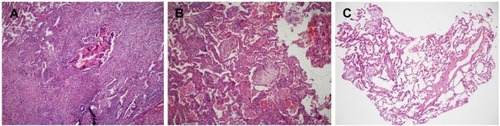
Table 1 Comparison of clinical, radiological, and PFT findings between COP and SOP cases
Table 2 Comparison of clinical, radiological, and PFT findings between cases diagnosed by surgical lung biopsy and by bronchoscopic biopsy
Table 3 Features of successful bronchoscopy group and failed bronchoscopy group