Figures & data
Figure 1 (A) The Western blot study suggested that retinal NGB expression in untreated eyes was significantly lower than in normal controls. (B) The retinal NGB level in the treated eyes was significantly higher than in normal controls. The retinal NGB level in the treated eyes was significantly higher than in untreated eyes, suggesting the AAV vector mediated pronounced NGB overexpression in the treated eyes. (C) The mRNA level of NGB in untreated eyes was significantly lower than that in normal controls. However, the mRNA level of NGB in the treated eyes was significantly higher than in normal controls. All values are presented as mean ± SD; #P<0.01 for differences compared between eye groups.
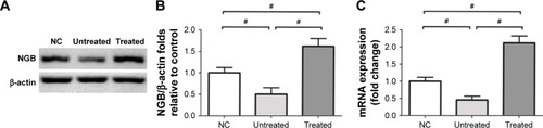
Figure 2 (A) The visual acuity and contrast sensitivity of untreated eyes were significantly lower than in normal controls, whereas visual acuity and contrast sensitivity of the treated eyes were both significantly higher than in the untreated eyes. Meanwhile, visual acuity and the contrast sensitivity of the treated eyes were significantly lower than in normal controls. (B) Representative ERG waveforms of different eye groups. (C) The photopic and scotopic b-wave amplitudes of untreated eyes were significantly smaller than in normal controls. Both photopic and scotopic b-wave amplitudes of treated eyes were significantly larger than those of the untreated eyes, suggesting NGB overexpression could ameliorate MNU-induced visual functional impairments. All values are presented as mean ± SD; *P<0.05 for differences compared between eye groups; #P<0.01 for differences compared between eye groups.
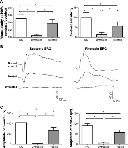
Figure 3 (A) Experimental animals were subjected to SD-OCT examination and their retinal thicknesses were quantified in vivo. Retinal thickness of the treated eyes was significantly smaller than that of normal controls. However, the retinal thickness of the treated eyes was significantly larger than that of untreated eyes. (B) The ONL of the untreated eyes disappeared after MNU administration. Conversely, a large proportion of ONL was retained in the retinas of treated eyes. The average ONL thickness of the treated eyes was significantly smaller than that of normal controls. The average ONL thickness of the treated eyes was significantly larger than that of the untreated eyes. All values are presented as mean ± SD; #P<0.01 for differences compared between eye groups.
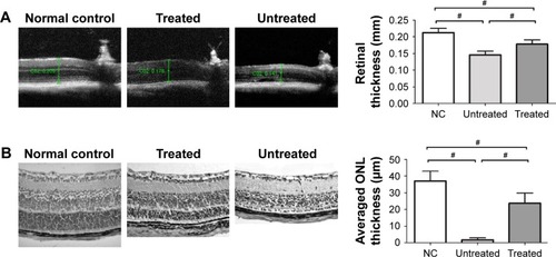
Figure 4 (A) The typical PNA staining was detected in the ONL of normal controls. However, no PNA staining was found in the retinas of untreated eyes. A substantial portion of PNA staining was detected in the retinas of the treated eyes. Both M- and S-opsin staining was detected throughout the retina of the treated eyes, although with a relatively decayed manner as compared with normal controls. (B) Retinal MDA concentration of the treated eyes was significantly lower than those of untreated eyes. The retinal SOD level in treated eyes was significantly higher than in untreated eyes, suggesting that the oxidative stress in the MNU-administered eyes was alleviated by NGB treatment. All values are presented as mean ± SD; #P<0.01 for differences compared between eye groups.
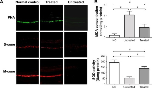
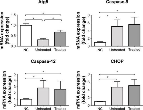
Figure 5 The retinal mRNA levels of bax, calpain-2, and caspase-3 in the treated eyes were significantly lower than in untreated eyes. On the other hand, the mRNA level of bcl-2 in the treated eyes was significantly higher than in untreated eyes. In untreated eyes, mRNA levels of CHOP, caspase-9, and caspase-12 were significantly higher than in normal controls. Expression levels of these ER stress sensors in the untreated eyes were not significantly different from those in the treated eyes. The mRNA level of Atg5 in untreated eyes was significantly lower than in normal controls. However, the mRNA level of Atg5 in treated eyes was significantly higher than in untreated eyes. All values are presented as mean ± SD; *P<0.05 for differences compared between eye groups; #P<0.01 for differences compared between eye groups.