Figures & data
Figure 1 A picture of the patient’s right hand.
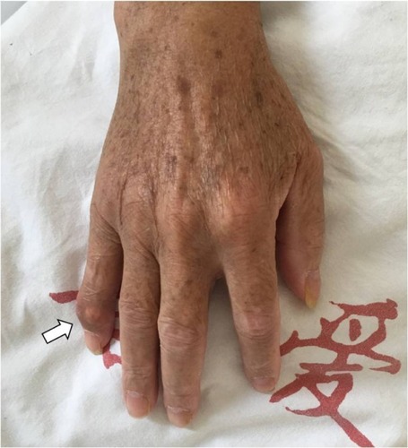
Figure 2 Plain X-ray films of the lumbar spine showing no obvious abnormalities except slight degenerative lumbar scoliosis (anteroposterior view on the left and lateral view on the right).
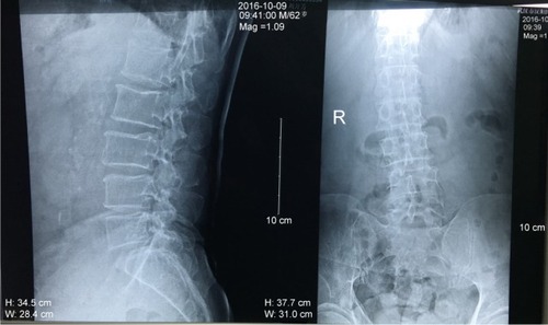
Figure 3 Computed tomography (CT) images of the lower spine.
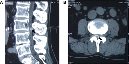
Figure 4 Lumbar spine magnetic resonance imaging (MRI) images demonstrating an abnormal round epidural collection at the L3/4 level, compromising the spinal canal and causing cauda equina compression (indicated by the arrows).
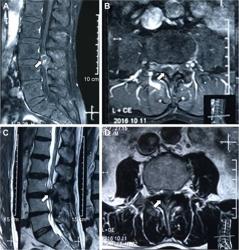
Figure 5 Intraoperative pictures showing white chalky material deposited in the epidural space of the posterior and lateral spinal canal (A). This material was partially encapsulated by fibrous tissue and grossly infiltrated the bone and soft tissue in several areas, which was removed completely with a curette (B and C).
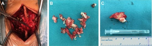
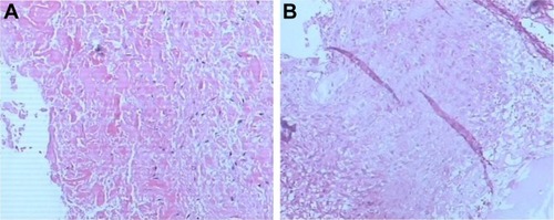
Figure 6 Microscopic examination of the specimen showed granulomatous tissue with structureless coagulative necrosis and fibrinoid necrosis (A and B).