
Figures & data
Figure 1 Flow chart of PABMT for delayed bone union in children.
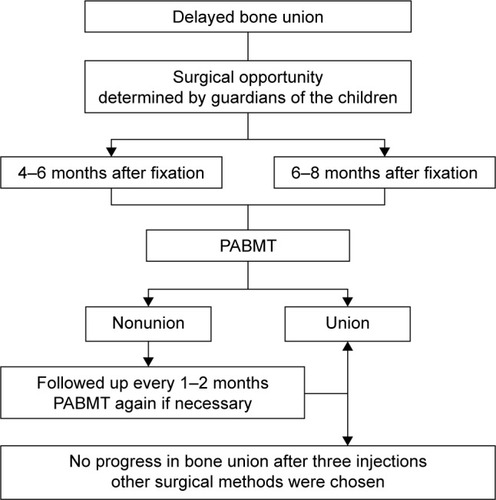
Table 1 The inclusion criteria and exclusion criteria
Figure 2 X-ray images of femoral fracture treated by external fixation and PABMT. A 12-year-old girl sustained a femoral shaft fracture in a traffic accident and was treated with external fixation. The frontal and lateral X-ray films showed clear fracture line with a small amount of callus 4 months after fixation. The girl was included in this study, and autologous bone marrow was injected to the delayed union site (A). One month later, there were obvious bony calluses, but the fracture line was visible (B). In the next 2 months, the patient underwent bone healing, and the external fixator was removed (C).
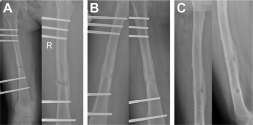
Figure 3 X-ray images of ulnoradial diaphyses fracture treated by internal fixation and PABMT. A 6-year-old boy suffered from ulnoradial diaphyses fracture following a fall and was treated with K-wire fixation (A). Three months after the operation, the ulnar fracture was healed clinically, but the fracture line in the radial shaft was clear without close endings (a sign of nonunion) (B). The K-wire in the ulna was pulled out, and the K-wire in the radius was left to continue to fix. Four weeks later, there was no progress in fracture healing – only a small amount of callus occurred and the fracture line was clear – the PABMT was carried out (C). At the sixth week after PABMT operation, the fracture line was fuzzy (D), the internal fixator was removed, and the patient underwent bone healing.
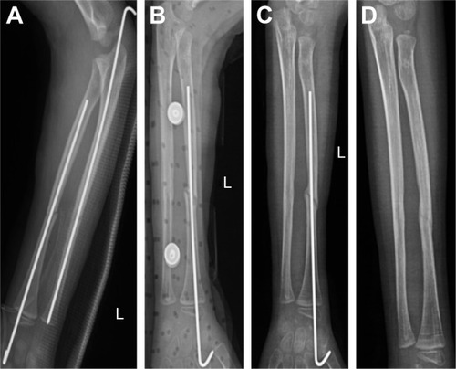
Table 2 Results of PABMT depending on transplantation period