
Figures & data
Figure 1 A 51-year-old female with esophagotracheal fistula for 7 months underwent division of the fistulous tract and closure of the trachea and esophagus.
Notes: (A) The tissue flap (arrow) is toward the trachea and before repair the entire opening of the fistula is opened toward the esophagus (tissue on the side of the trachea is scant). (B) The opening (between the vascular clamps) of the fistula is occluded with the tissue flap. Because the tissue flap is pulled toward the trachea, the gastric tube in the esophagus is seen. (C) The opening of the fistula after occlusion with the tissue flap.
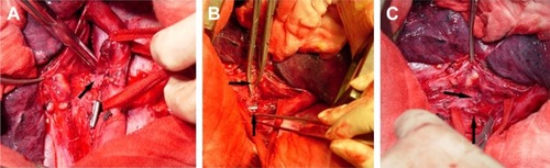
Table 1 Demographic and baseline information of patients (N=14)
Figure 2 Preoperative diagnostic investigation.
Notes: (A) Esophagography demonstrates the outflow of radiopaque diagnostic medium from the esophagus into the trachea in a 51-year-old female with esophagotracheal fistula. (B) CT scan shows the opening of the esophagotracheal fistula. (C) Gastroendoscopy reveals the opening of the fistula in the esophagus in the same patient.
Abbreviation: CT, computed tomography.
Abbreviation: CT, computed tomography.
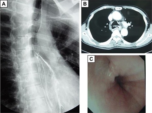
Figure 3 The location of the esophagotracheal fistula is determined during the operation.
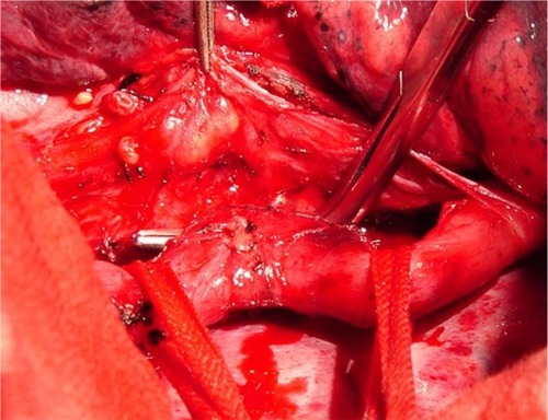
Table 2 Surgical treatments and outcomes of patients with acquired benign esophagorespiratory fistulae in the literature