Figures & data
Figure 1 Age distribution of multiply injured patients without blunt injury of the cervical arteries (control), with CAD, and with VAD.
Abbreviations: CAD, carotid artery dissection; VAD, vertebral artery dissection.
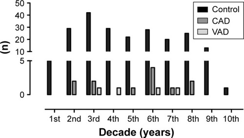
Table 1 Data on patients, injury severity, and mortality
Figure 2 Trauma mechanism of multiply injured patients without blunt injury of the cervical arteries (control), with CAD, and with VAD.
Abbreviations: CAD, carotid artery dissection; VAD, vertebral artery dissection.
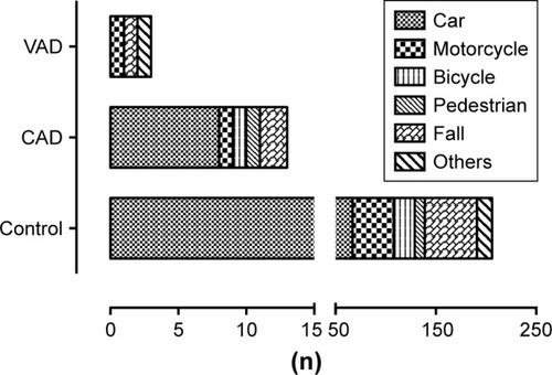
Figure 3 Trauma severity measured by ISS of multiply injured patients without blunt injury of the cervical arteries (control), with CAD, and with VAD.
Note: Values are shown as mean ± SD.
Abbreviations: CAD, carotid artery dissection; ISS, Injury Severity Score; VAD, vertebral artery dissection.
Abbreviations: CAD, carotid artery dissection; ISS, Injury Severity Score; VAD, vertebral artery dissection.
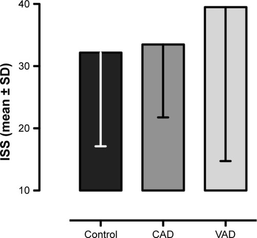
Table 2 Concomitant injuries
Figure 4 Image of traumatic carotid artery dissection on the right side.
Notes: (A) Axial polytrauma CT clearly depicts the dissection membrane in the right internal carotid artery (arrow). (B) No flow (arrow) can be seen distal of the CAD right before entering of the carotid artery into the petrous bone. (C) PD fs axial MRI sequence, acquired 1 week later, shows the right-sided CAD revealing the hyperdense carotid wall hematoma (arrow).
Abbreviations: CAD, carotid artery dissection; CT, computed tomography; MRI, magnetic resonance imaging; PD fs axial, axial fat-saturated proton density.
Abbreviations: CAD, carotid artery dissection; CT, computed tomography; MRI, magnetic resonance imaging; PD fs axial, axial fat-saturated proton density.
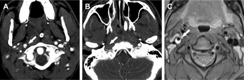
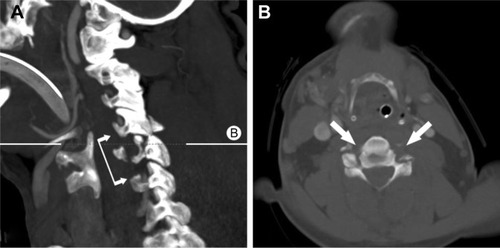
Figure 5 Bilateral vertebral artery dissection in cervical C5/6 facet fractures with dislocation.
Notes: (A) No flow in the vertebral arteries from C5 to C7 (connected arrows). B delineates plane of axial CT image B. (B) Axial CT image shows no flow in both vertebral arteries (arrows).
Abbreviation: CT, computed tomography.
Abbreviation: CT, computed tomography.