Figures & data
Figure 1 Distribution (in %) of localizations of infected nonunions.
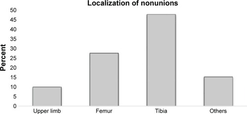
Figure 2 Distribution (in %) of fracture types causing infected nonunions.
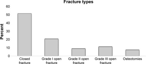
Table 1 Detected species from tissue samples of all 171 infected nonunions with positive microbial evidence
Figure 3 Successful nonunion treatment after Masquelet technique.
Notes: Nonunion of the distal tibia 18 months after fracture with microbiological evidence of coagulase-negative Staphylococcus spp. (A). Postoperative image after changing the osteosynthetic material together with extensive debridement and implantation of a bone cement spacer according to Masquelet technique step 1 (B). Final image after Masquelet step 2 with spacer removal, metaphyseal locking compression plate re-osteosynthesis and implantation of cancellous bone of the femur into the fracture gap. The patient showed complete fracture healing after 16 months (C).

Figure 4 Unsuccessful nonunion treatment after Masquelet technique.
Notes: Nonunion of the distal tibia 20 months after fracture with microbiological evidence of a mixed infection (A). Postoperative image after changing the tibial nail and extensive debridement with implantation of a bone cement spacer according to Masquelet step 1 and additional implantation of a locking compression plate at the fibula (B). Masquelet step 2 was performed 6 weeks later with spacer removal and re-osteosynthesis using an Expert Tibial Nail PROtect® at the tibia together with cancellous bone. At 22 months after Masquelet step 2, the patient did not show fracture healing in X-rays (C) and computed tomography scan (D), yet.
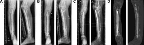
Figure 5 Fusion rates (in %) of cases with infection with the 3 most frequently detected bacteria and cases without detected microbial infection compared to all 171 cases of infected nonunion with positive microbial evidence.
Note: Lower part of each row shows complete bone fusion rate with absent fracture line, upper part depicts rate of incomplete bone fusion with partial fracture line.
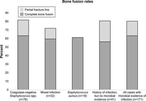
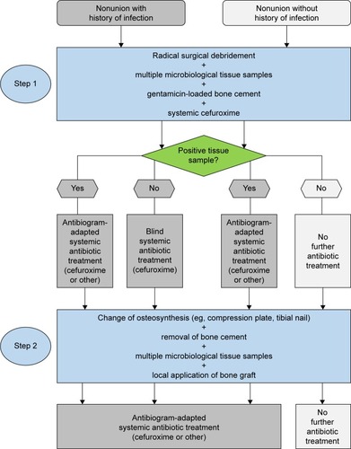
Figure 6 Schematic algorithm of Masquelet step 1 and 2 with peri- and postoperative antibiotic treatment.