Figures & data
Figure 1 Imaging for Cases 1 and 2.
Notes: (A) Magnetic resonance imaging of the abdomen on presentation of Case 1. Multiple cystic lesions were seen with short T1 and long or short T2 signals (white arrow); left kidney was pushed and compressed by the mass (yellow arrow). (B) Computed tomography scan shows a huge mass from the left side of retroperitoneal to iliac fossa (yellow arrow) in Case 2; the functional solitary left kidney was pushed and compressed by the mass (white arrow).
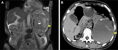
Table 1 Laboratory test results before surgery
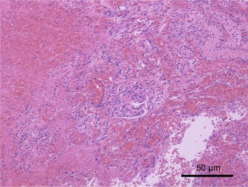
Figure 2 Pathologic findings of Case 1.
Notes: The majority of mass consisted of blood clots and fibrinous exudate cells, and a few broken and necrotic glomerular and renal tubules were seen. No evidence of malignancy and no bacteria were found in the necrotic foci. PAS and D-PAS were negative. Magnification ×100.
Abbreviations: PAS, periodic acid-Schiff; D-PAS, D-periodic acid-Schiff stain.
Abbreviations: PAS, periodic acid-Schiff; D-PAS, D-periodic acid-Schiff stain.