
Figures & data
Figure 1 Preoperative imaging diagnoses. (A) Chest CT scan at the day before admission shows a smooth cystic tumor measuring 67 × 51 × 60 mm (white arrow). CT value was 28 HU. The esophagus was compressed by the cystic tumor. (B) An abdominal CT at the day before admission revealed a normal shape of the stomach (white arrow). (C) Transverse contrast-enhanced chest CT at 5 days after admission showed the sharply defined mass measuring 47 × 70 × 85 mm (white arrow). CT value was 31 HU. (D) Transverse contrast-enhanced abdominal CT at 5 days after admission showed a huge contusion of the stomach wall (black arrow).
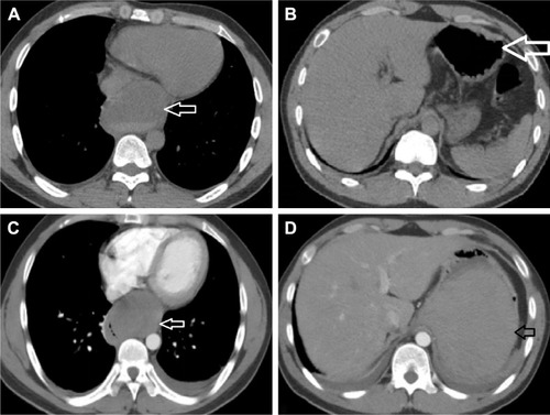
Figure 2 Gastroscopy images. (A, B) After admission, gastroscopy showed a mass with a smooth surface causing a partial obstruction of the lower esophagus and stomach fundus. (C, D) The day before admission, gastroscopy showed a mass with a smooth surface causing partial obstruction of the lower esophagus and a normal stomach fundus.
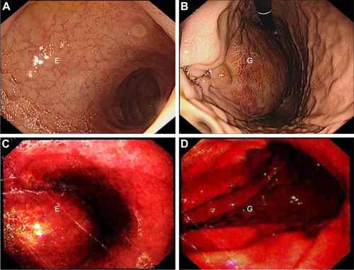
Figure 3 Intraoperative images. (A) Brown fluid was observed in the bronchogenic cyst after dissection of the cystic wall. (B) The nutrient artery (arrow) supplying the cyst was revealed distinctly. (C) The shape of the bronchogenic cyst was revealed. (D) Because of the gastric hematoma, a violet surface (arrow) was observed after incision of the diaphragm.
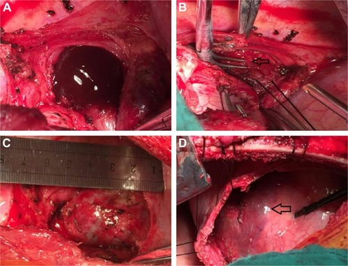
Figure 4 Immunohistochemical and histopathological examination of the cyst sections. (A) Positive expression of CA199, original magnification ×200. (B) Positive expression of CA125, original magnification ×200. (C, D) The cystic wall was lined with ciliated columnar epithelium. The wall also contained cartilage and bronchogenic glands (H&E stained, original magnification ×40).
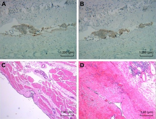
Figure 5 (A) A control esophagogram showed that the esophagus (black arrow) had neither stenosis nor leakage. (B) The shape and function of the stomach (white arrow) were normal.
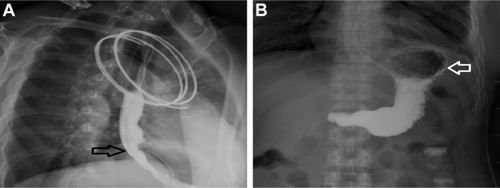
Table 1 Summary of published cases with periesophageal or intramural esophageal bronchogenic cysts