Figures & data
Figure 1 Preoperative and postoperative chest X-ray of the patient.
Notes: (A) Posterior–anterior chest X-ray preoperatively showing pulmonary edema and a mass in the left lung. (B) Chest X-ray 3 h postoperatively showing pulmonary edema was relieved, and the lung mass became distinct.
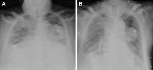
Figure 2 Hyperechoic lumpy mass detected on transthoracic echocardiography.
Note: Apical four chamber view showing the mass in the left atrium (orange arrow).
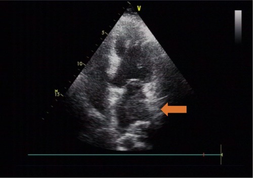
Table 1 Arterial blood gas analysis and ACT during surgery
Table 2 Vital signs and ventilation parameters during surgery
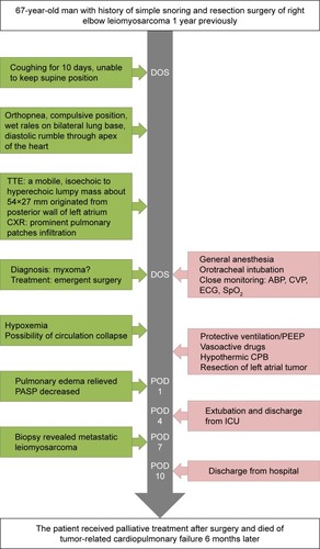
Figure 3 Timeline of interventions and outcomes.
Abbreviations: ABP, arterial blood pressure; CPB, cardiopulmonary bypass; CVP, central venous pressure; CXR, chest X-ray; DOS, day of surgery; ECG, electrocardiogram; ICU, intensive care unit; PASP, pulmonary arterial systolic pressure; PEEP, positive end expiratory pressure; POD, postoperative day; TTE, transthoracic echocardiography; SpO2, pulse oximetry.