Figures & data
Figure 1 Measurement of AP diameter of cervical canal and spinal cord compression on T2 sagittal MR imaging.
Notes: D1–D4 were diameters of cervical canal at the mid-vertebra level from C4 to C7. AP diameter of cervical canal (mm)=(D1+D2+D3+D4)/4. a and b are the spinal cord diameters of narrowest part and the C2/C3 intervertebral level. Spinal cord compression (%)=a/b.
Abbreviations: AP, anteroposterior; MR, magnetic resonance.
Abbreviations: AP, anteroposterior; MR, magnetic resonance.
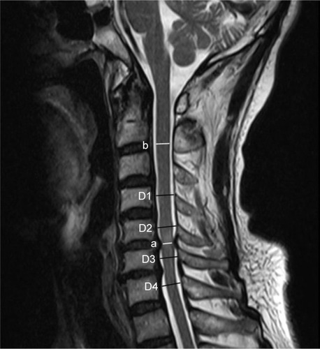
Figure 2 The lumbar canal was calculated by a ratio of the mid-sagittal spinal canal diameter at the level of the intervertebral disc to the spinal canal diameter at the mid-vertebra level of the upper vertebral body.
Note: The percentage of lumbar canal stenosis (%)=(a/A+b/B+c/C+d/D)/4.
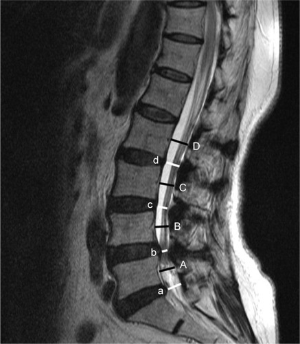
Table 1 Classification system for lumbar spinal stenosis based on MR images
Figure 3 Kaplan–Meier survivorship curve for symptomatic LCS after CSM surgery.
Abbreviations: CSM, cervical spondylotic myelopathy; LCS, lumbar canal stenosis.
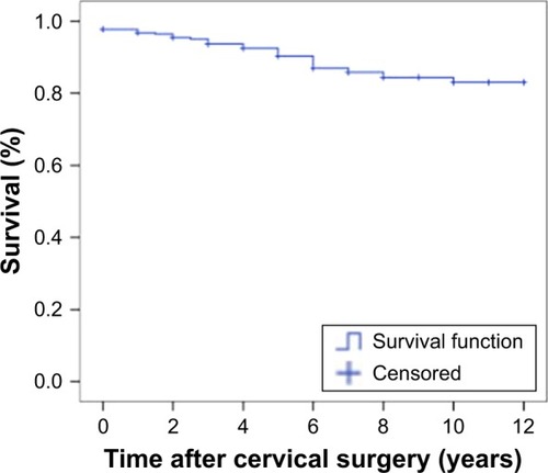
Table 2 Patients’ demographic and baseline data for two groups
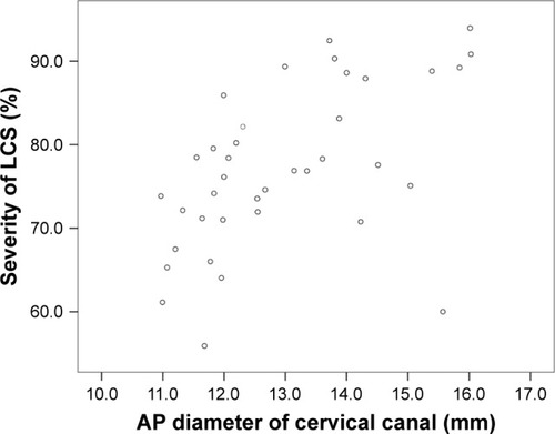
Figure 4 Scatter diagram showing the relationship between AP diameter of cervical canal and severity of LCS.
Abbreviations: AP, anteroposterior; LCS, lumbar canal stenosis.