
Figures & data
Figure 1 Evaluation of vessel diameter in CE-CT.
Notes: (A) Parasagittal curved MPR of a proximal TC stenosis with location of the orthogonal cuts at the normal (1) and maximally stenosized (2) lumen. Orthogonal planes where the measurements are performed are presented in (B and C). Degree of stenosis (s) in (%) is calculated using the formula s = (a−b)/a × 100.
Abbreviations: CE-CT, contrast enhanced-computed tomography; MPR, multiplanar reconstruction; TC, celiac trunk.
Abbreviations: CE-CT, contrast enhanced-computed tomography; MPR, multiplanar reconstruction; TC, celiac trunk.
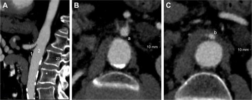
Table 1 Definitions used to grade calcifications of the supplying arteries of the gastric tube seen on preoperative CT images by van Rossum et alCitation9
Figure 2 Degree of (A) TC and (B) SMA stenosis in patients with and without anastomotic insufficiency after esophagectomy and gastric pull-up.
Note:
*Outliers with a distance from the box of more than 3xIQR.
Abbreviations: SMA, superior mesenteric artery; TC, celiac trunk.
Abbreviations: SMA, superior mesenteric artery; TC, celiac trunk.
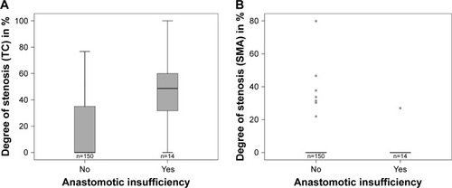
Table 2 Frequency of calcification score of patients with/without AI
Table 3 Histopathologic results and preoperative comorbidities in association with anastomotic leak