
Figures & data
Figure 1 Axial muscle MRI of thighs and legs and muscle biopsy findings (gastrocnemius).
Notes: (A–H) MRI of the thighs: axial T1-weighted images (A and B) show mild fatty degeneration of right and left semimembranosus (arrow). Axial fat-suppressed T2-weighted images (C and D) reveal edema of the right semimembranosus and very mild edema of left semimembranosus (arrow) and long head of biceps femoris (arrow head). MRI of the legs: axial T1-weighted images (E and F) show fatty degeneration of medial head of gastrocnemius (arrow) and soleus bilaterally (arrowhead). Axial fat-suppressed T2-weighted images (G and H) reveal mild edema of the medial head of gastrocnemius (arrow) and soleus(arrow head) bilaterally. (I–N) Histological findings: (I) H&E stain: variation in fiber size, connective tissue proliferation and necrosis. (J) Gomori trichrome: connective tissue proliferation. (K–N) Positive immunostaining for MHC class I CD4, CD8 and CD68, respectively. (I–N) 10×; scale bar: 1 μm.
Abbreviations: MHC, major histocompatibility complex; MRI, magnetic resonance imaging.
Abbreviations: MHC, major histocompatibility complex; MRI, magnetic resonance imaging.
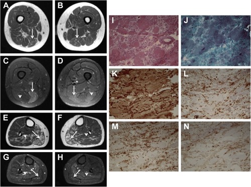
Figure 2 Axial muscle MRI of thighs and legs.
Notes: Thighs (A–D): diffuse edema of the right semimembranosus and tenuous edema of the left semitendinosus and femoral biceps. Legs (E–H): diffuse edema and discreet fatty infiltration of the soleus muscle and the medial and lateral heads of both gastrocnemii.
Abbreviation: MRI, magnetic resonance imaging.
Abbreviation: MRI, magnetic resonance imaging.
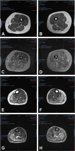
Table 1 Previous cases reported with anti-HMGCR myopathy simulating dystrophic-like pattern