
Figures & data
Figure 1 The abdominal computed tomography (CT) scan confirmed a well-defined cystic neoplasm in the pancreatic tail (A), without enhancement in the arterial phase (B) and the portal phase (C). Endoscopic ultrasonography (EUS) showed a 3.5 cm multilocular cystic lesion in the pancreatic tail with an internal nodule (D).
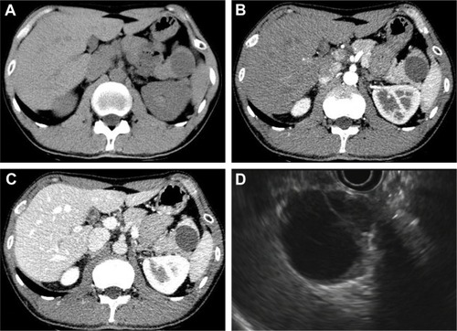
Figure 2 (A) Gross appearance of the epidermoid cyst in an intrapancreatic accessory spleen (ECIPAS), with 4 cm at its greatest diameter. (B) Microscopic analysis revealed a multilocular cyst surrounded by accessory splenic tissue in the pancreas parenchyma, and the cyst wall showed a thin multilayered squamous epithelium (H&E staining, ×50).
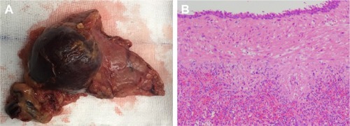
Table 1 Reported studies of an ECIPAS in the English language literature