Figures & data
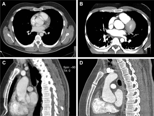
Figure 1 (A) and (C) showing non-ECG-gated prereferral CTA suggesting aortic dissection in patient 1; (B) and (D) showing postoperative scan acquired in gated high-pitch mode and with subsecond level acquisition time.
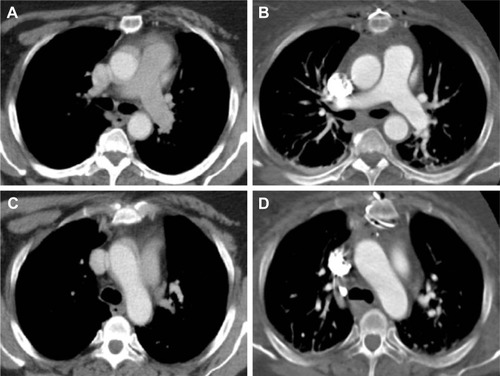
Table 1 Laboratory findings at admission
Figure 2 (A) and (C) showing non-ECG-gated prereferral CTA suggesting circumferential dissection of the ascending aorta in patient 2; (B) and (D) showing gated repeat CTA without misleading artifacts.
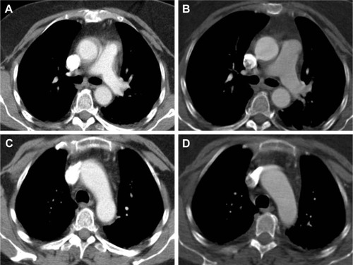
Figure 3 (A) Non-ECG-gated prereferral CTA suggessting dissection of the ascending and descending aorta and (B) ECG-gated repeat CTA confirming only type B dissection in patient 4. (C) Suspicious structures at the level of the proximal ascending aorta in the non-ECG-gated prereferral scan of patient 3 that are absent in the ECG-gated repeat scan shown in (D).
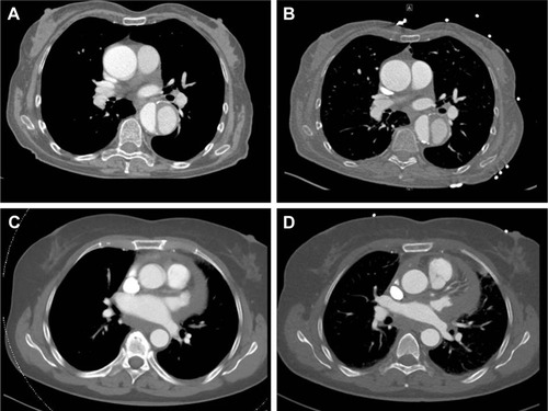
Figure 4 (A) and (C) showing non-ECG-gated axial and coronal CTA images suggesting dissection at the level of the aortic root in patient 5. (B) and (D) showing the ECG-gated CT scan of the same patient subsequently performed on a 256-MSCT device that showed no aortic dissection.