Figures & data
Figure 1 Color photography showing penetrating keratoplasty with peripheral opacification.
Note: There is a clinically visible anterior synechiae from 3 to 6 o’clock.
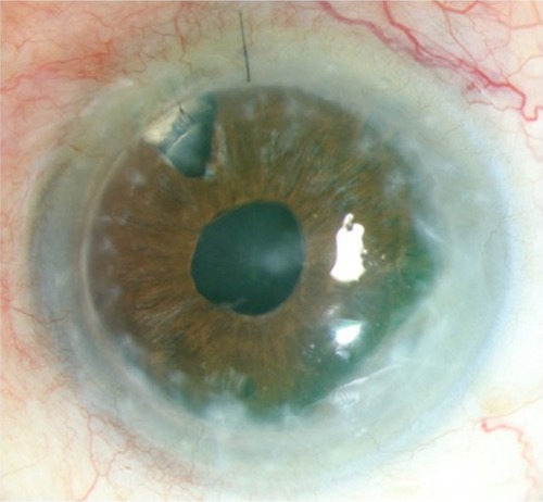
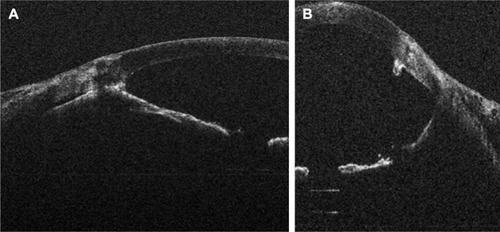
Figure 2 Intraoperative OCT showing (A) peripheral anterior synechiae posterior to the opacification and (B) after surgery, deepening of the anterior chamber and lysis of adhesion.
Abbreviation: OCT, optical coherence tomography.