
Figures & data
Figure 1 Abdominal CT showing the gas-liquid levels and dilatation of the intestinal tract and an obscured appendiceal area revealing edema of this area (circle).
Note: However, when we reviewed the CT repeatedly after the operation, the images appeared to show that the appendix was shaped like a horseshoe, a finding that was easily ignored.
Abbreviation: CT, computed tomography.
Abbreviation: CT, computed tomography.
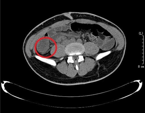
Figure 2 The horseshoe appendix was bundled and cut into two parts (arrow) to manage it side by side for severe adhesions in the appendiceal area.
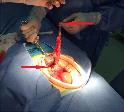
Figure 3 Both divided bases (arrow) are completely shown.
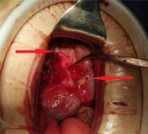
Figure 4 Frontally displaced mesentery (our case is of this type).
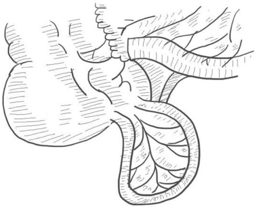
Figure 5 Sagittally displaced mesentery.
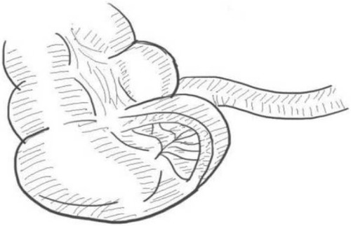
Table 1 Cases of a horseshoe appendix
Table 2 Classification of appendiceal anomalies