Figures & data
Figure 1 Preoperative 3D-CT.
Abbreviations: 3D-CT, three-dimensional computed tomography.
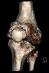
Figure 2 Preoperative full-length, weight-bearing lower extremity anteroposterior view.
Figure 3 Intraoperative radiograph showing the intra-articular bone resections made parallelly to the preexisting joint obliquity (white arrows), osteotomized tibial tubercle (gray arrows), and insertion of Kirschner wire in the area of the planned tibial osteotomy (black arrow).
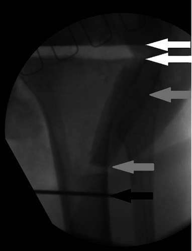
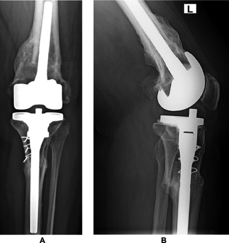
Figure 4 Postoperative radiograph taken at 5-year follow-up. (A) Anteroposterior view and (B) lateral view.