Figures & data
Figure 1 Measurements of RPS in 2nd, 5th and 7th cervical spine level in median sagittal MRI images.
Notes: The thickness of RPS (solid double arrow) is measured by the antero-posterior distance between the posterior margin of pharynx, larynx or trachea and the anterior bony margin of cervical vertebra. The proportion of RPS to vertebral body was calculated as a ratio of the thickness of RPS to A–P diameter of each vertebral body (dotted double arrow) multiplied by 100.
Abbreviations: A–P, antero-posterior; MRI, magnetic resonance imaging; RPS, retropharyngeal space.
Abbreviations: A–P, antero-posterior; MRI, magnetic resonance imaging; RPS, retropharyngeal space.
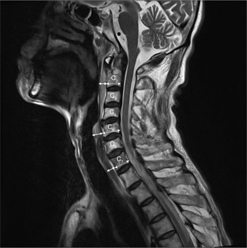
Table 1 Characteristics of patients
Table 2 Quantitative measurement of retropharyngeal space between easy and difficult laryngoscopy during anesthesia induction in patients with traumatic cervical spine injury
Table 3 Univariate logistic regression analysis for the prediction of difficult laryngoscopy in patients with traumatic cervical spine injury
Figure 2 ROC curve and AUC of the RPS at the 2nd cervical spine level.
Notes: The areas under the receiver operator characteristic curve are 0.97 (95% CI: 0.94–1.01; p<0.001) and 0.96 (95% CI: 0.92–1.01; p<0.001), respectively. Cutoff values of RPS thickness and the proportion of RPS to the 2nd cervical vertebral body are 7.94 mm and 48.4% (p<0.001 and p<0.001, respectively).
Abbreviations: AUC, area under the curve; ROC, receiver operating characteristic; RPS, retropharyngeal space.
Abbreviations: AUC, area under the curve; ROC, receiver operating characteristic; RPS, retropharyngeal space.
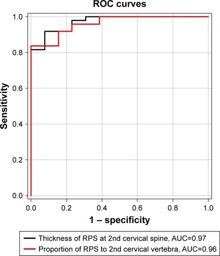
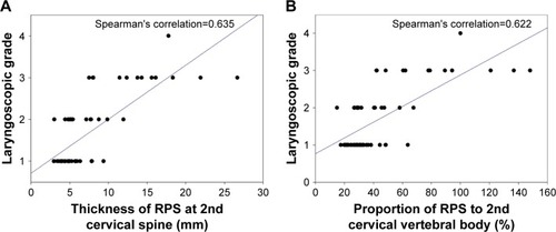
Figure 3 The relationships between laryngoscopic grade using Cormack–Lehane classification and (A) RPS thickness at the 2nd cervical spine level, and (B) proportion of RPS to A–P diameter of the 2nd cervical vertebral body.
Abbreviations: A–P, antero-posterior; RPS, retropharyngeal space.