Figures & data
Table 1 Demographics and symptoms of 7 patients
Figure 1 Port placement for retroperitoneal robot-assisted laparoscopic upper pole heminephrectomy. (A) Trocar locations. Two 12-mm ports were placed and used as a camera port and an assistant port. Two 8-mm ports were placed for the remaining robotic arms. Open circles represent 8-mm ports, and closed circles 12-mm ports. (B) Patient positioning for left dissection.
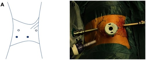
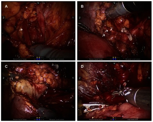
Figure 2 Surgical video screenshots of the retroperitoneal robot-assisted laparoscopic upper pole heminephrectomy. (A) The dilated upper (black arrow) and lower (white arrow) ureters were clearly identified. (B) The upper pole vessels (white arrow) were dissected free, ligated using Hem-o-lok clips. (C) By cranial traction of the proximal ureteral stump, dissection proceeded along the demarcation line between the upper and lower pole. (D) Renorrhaphy was performed.