
Figures & data
Figure 1 Shoulder dislocation in Patient 2. A normal glenoid cavity conforms to the red circle. The anterior edge of the glenoid cavity is straightened (white arrow) indicating bone defect, at the 2:00–6:00 position (A). The humeral head (yellow arrow) is displaced downward (B). The humeral head has been repositioned and returned to its original position (C).
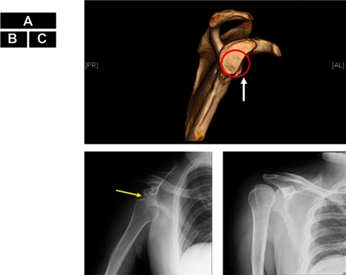
Figure 2 MRI pre- and post-corpus callosotomy for Patient 1. Preoperative coronal (A), sagittal (B), postoperative coronal (C) and sagittal (D) MRI FLAIR of Patient 1.fi. The arrows show that the corpus callosum is totally removed (B and D).
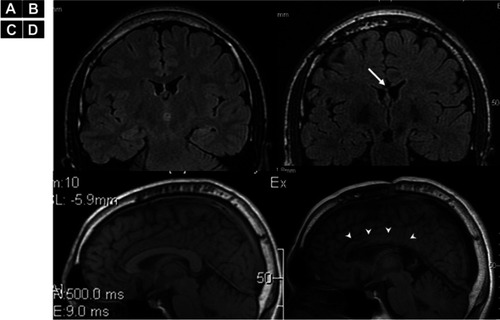
Figure 3 Electro Corticography (ECoG) of Pre- and post-corpus callosotomy for Patient 2. (A) Pre-corpus callosotomy ECoG monitoring from bilateral frontal lobe cortices shows generalized poly-spikes. (B) Post-corpus callosotomy ECoG shows reduced generalized poly-spikes lateralized to the left hemisphere. The epileptiform discharges on the right side are eleminated (arrow). As scalp EEG showed epileptiform discharges predominantly in the left hemisphere, we used an 8-contact strip on the left hemisphere and a 6-contact strip on the right hemisphere.
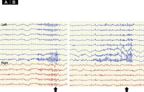
Table 1 Clinical information and data between pre- and post-callosotomy