
Figures & data
Figure 1 A chest X-ray revealed patchy consolidation in the bilateral upper and left lower lung field (white arrow).
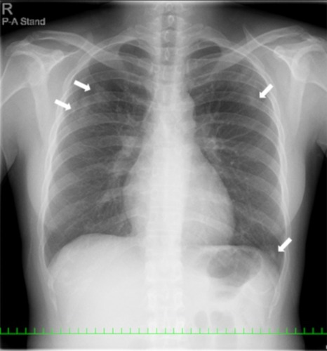
Figure 2 CT scans showed bilateral patchy consolidation surrounded by ground-glass opacity (white arrow).
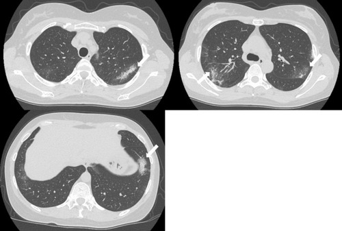
Figure 3 BAL fluid showed increased numbers of eosinophils with Papanicolaou staining (A, ×400). Under high magnification, the eosinophils were stained light green with nuclear localization (B, ×800).
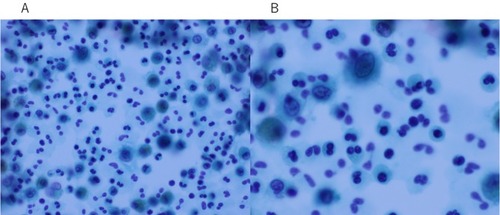
Figure 4 A chest X-ray and chest CT images showed an improvement in lung infiltrates after the discontinuation of natalizumab.
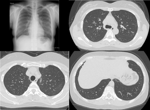
Figure 5 The clinical course of the patient.
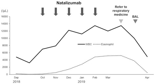
Table 1 Laboratory Findings On The Initial Visit
Table 2 Summary Of Cases With Respiratory Adverse Events Associated With Natalizumab