
Figures & data
Table 1 A Comparison of the Preoperative General Condition Between the Two Groups
Figure 1 A 56-year-old female patient presented with numbness and weakness in both lower limbs for 19 months. (A, B) Preoperative computed tomography (CT) showed TOLF at the level of T3–5, and ossified ligamentum flavum in fused-type. (C) Magnetic resonance imaging (MRI) showed that the low-signal ossification invaded the spinal canal from the rear, and the spinal cord was severely compressed. (D) The lamina was clamped using a towel clamp, and a bone pry was inserted into the gap at the edge of the free lamina. (E, F) The laminae and ossified ligamentum flavum were excised from the head to the tail. (G, H) Postoperative CT showed that the majority of the vertebral lamina and ossification were removed, and the spinal canal was unobstructed. (I) Postoperative MRI showed disappearance of the compression behind the spinal cord.
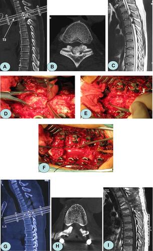
Figure 2 A 58-year-old female patient presented with a walking disability in both of the lower limbs for 17 months. (A) Preoperative X-ray showed that the thoracic vertebrae degenerated, and the local Cobb angle was 8.5°. (B, C) Preoperative magnetic resonance imaging and computed tomography showed ossification of the ligamentum flavum at the level of T9–12, and the spinal canal demonstrated severe stenosis. (D, E) A diagram of lamina osteotomy. (F, G) The decompression range and imaging findings after laminae replantation. (H, I) The ossification was thoroughly removed, and the spinal canal was unobstructed. (J) One year after surgery, the local Cobb angle increased to 11.4°.
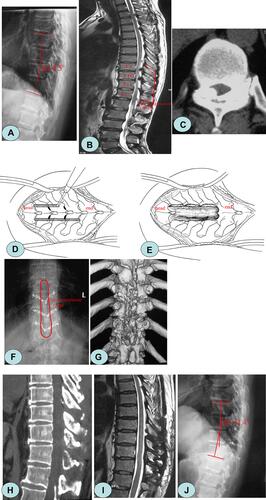
Table 2 Comparison of Intraoperative Data and Complications Between the Two Groups
Table 3 Changes in JOA Score and Cobb Angle Before and After Surgery in the Two Groups
Table 4 Comparison of ASIA Grading Before and After Surgery in the Two Groups (n)