
Figures & data
Figure 1 Using customized software (Mimics 14.01 software, Materialise, Leuven, Belgium), 3-D models of the affected and contralateral normal humeri were constructed (A). On the affected humerus and the mirror image of the contralateral normal humerus, the centers of the capitellum and trochlea were obtained with the use of a circle-fit algorithm at the lateral surface of the capitellum, and the center (flexion-extension) axis was created as a line through the geometric centers of the trochlea and capitellum (B). Then, the distal part of the model of the affected humerus was superimposed onto the corresponding part of the mirror image of the normal humerus to measure the degrees of varus and extension components of the deformity by point and surface registration using the medial and lateral epicondyles, the distal articular surface, and the flexion-extension axis as references (C).
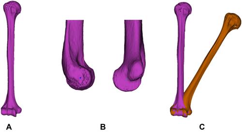
Figure 2 In three-dimensional measurements of cubitus varus, the anteroposterior view of the varus deformity angle (A) was obtained by measuring the angle between the humeral axes of the affected and normal humeri. Lateral view of the extension deformity angle (B). The internal rotation deformity angle was measured as the difference in retroversion angles (θ) of the affected and normal humeri (C).
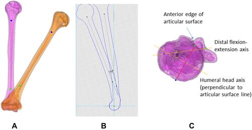
Figure 3 In radiographic measurements, the humerus-elbow-wrist angle (A) comprised the angle between the humeral axis and a line passing through the proximal and distal midpoints of the radius and ulna. The tilting angle (B) was determined by the anterior tilt of the articular condyles with respect to the humeral axis on a lateral radiograph. For physical measurements, the carrying angle (C), elbow flexion (D), and shoulder internal rotation (E) were estimated.
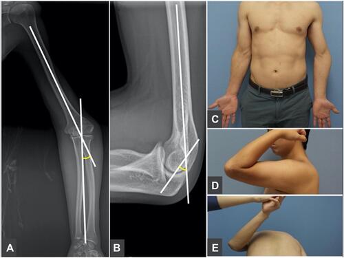
Table 1 Comparison of Measurements for Each Component of Cubitus Varus