Figures & data
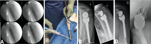
Figure 1 Implant coating of a plate; (A) tibial defect after stabilization with a LCP; (B) Augmentation of the tibial defect and the plate; (C) after closing the defect with a ALT-flap; (D) bone healing in CT-scan 15 month later.
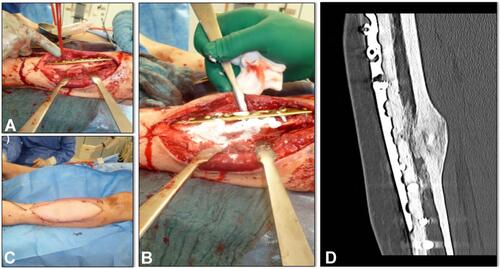
Figure 2 Implant coating of a nail. (A) filling up the intramedullary canal with Cerament V and implantation of the augmented nail; (B) coating of the nail before implantation; (C) x-ray 22 moths after implant coating.
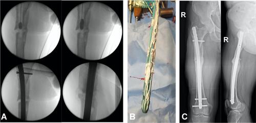
Figure 3 : (A) + (B) Removal of the loosened humeral nail; (C) Implantation of a cement spacer.
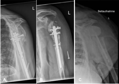
Figure 4 Implant coating of a shoulder prosthesis; (A) Filling the medullary canal with 19 ml Cerament V; (B) coating of the prosthesis with Cerament V; (C) x-ray 2 moths after implant coating; (D) x-ray 15 moths after implant coating.