
Figures & data
Table 1 Preoperative General Conditions of the Two Groups
Figure 1 (A–F) depicts a 45-year-old male patient who was administered general anaesthesia before undergoing open reduction, internal fixation, and bone graft for the treatment of his left tibial plateau fracture. During the surgery, trapdoor-procedure-based bone harvesting was used. (A) illustrates the location of the bone harvesting site prior to surgery. (B) displays the site after the trapdoor was opened. (C) depicts the site when the trapdoor was placed back to the iliac spine. (D) represents a post-operative image of the pelvis. (E) illustrates a clear left tibial plateau fracture and articular surface collapse. (F) illustrates a post-operative left tibial plateau.
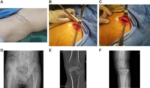
Figure 2 (A–E) depicts a 53-year-old female patient who was administered general anesthesia before undergoing open reduction, internal fixation, and bone grafting to treat her right tibial plateau fracture. During surgery, tricortical iliac bone harvesting was used. (A) illustrates the location of the bone-harvesting site before surgery. (B) displays the bones being harvested. (C) shows a postoperative image of the pelvis. (D) presents a clear right tibial plateau fracture and articular surface collapse. (E) is a postoperative image of the tibial plateau fracture.
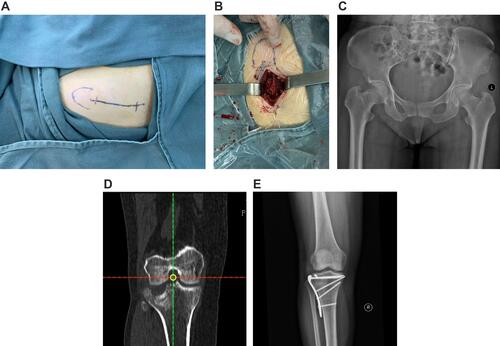
Table 2 Iliac Bone-Graft Donor Site Pain Scores (x ± S; Measured Using SF-MPQ-2) of the Two Patient Groups
Table 3 Differences in Iliac Bone-Graft Donor Site Incision Length, Intraoperative Blood Loss, Amount of Bones Harvested, Operation Time, and Incidence of Complications Between the Two Patient Groups