
Figures & data
Figure 1 Computed tomography revealing intracranial pneumocephalus, evident at the top of the forehead, and compression of the bilateral frontal lobes.
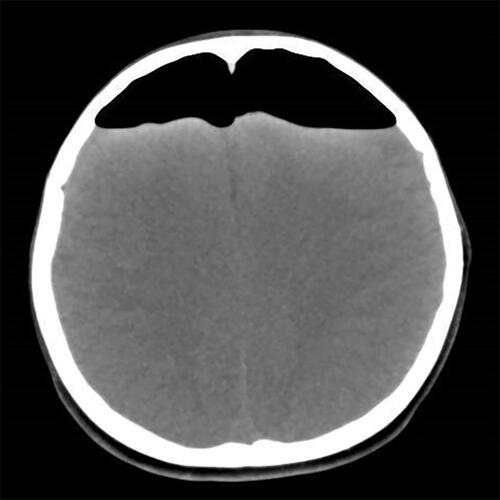
Figure 2 Magnetic resonance imaging revealing pneumocephalus in the bilateral ventricles, with slight enlargement of the anterior horn of the lateral ventricles.
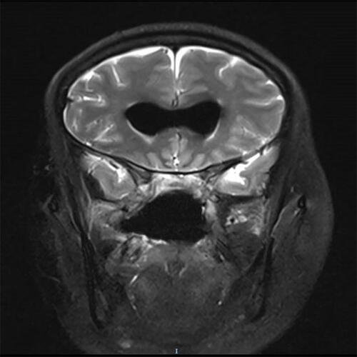
Figure 3 Magnetic resonance imaging revealing a significant decrease in bilateral ventricular pneumocephalus.
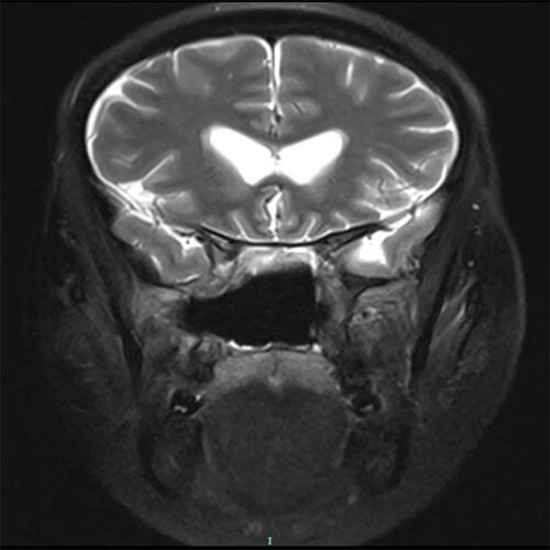
Figure 4 Computed tomography revealing obvious pneumocephalus in the intracranial lobe, with compression of the brain tissue, and marked enlargement of the ventricles and fissures.
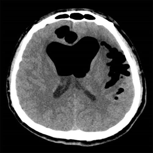
Figure 5 Computed tomography revealing a large area of pneumocephalus in the bilateral anterior cranial fossa. The frontal retention of air caused widening of the interhemispheric fissure, leading to a peaked appearance of the frontal poles, commonly referred to as “Mount Fuji sign”.
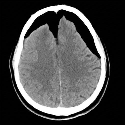
Table 1 Summary of All Reported Cases of TP Caused by Endoscopic Endonasal Surgery