Figures & data
Figure 1 Clinical images of ARPC. Scattered keratotic papules on the trunk, part of the lesions may have umbilical recesses, and the shape is crater-shaped (White arrow).
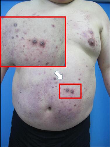
Figure 2 Clinical images of ARPC. (A) Koebner Phenomenon due to scratching can be seen at the elbow, and the lesions are fused into a piece (Yellow arrow). Typical rash for crater-shaped (White arrow); (B) crater-shaped rash of waist; (C) Koebner Phenomenon of lower limb.
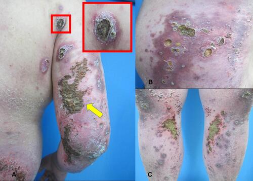
Figure 3 Histopathology of ARPC. (A) collagen fibers that vertically penetrated the epidermis (White arrow, HE×40); (B) HE×100; (C) the collagen fibers of the epidermis are stained light blue (Masson stain×400).
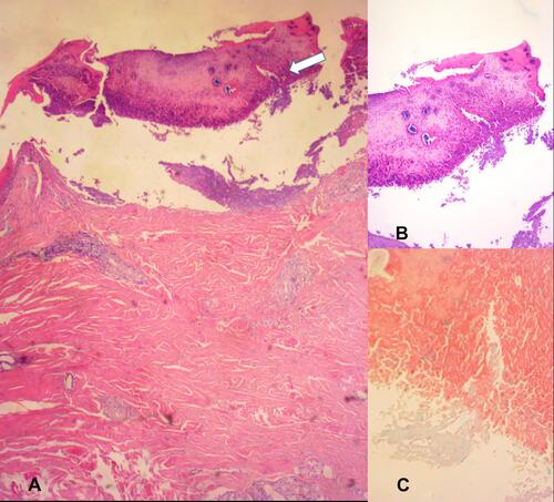
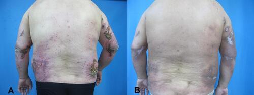
Figure 4 Clinical images before and after ARPC treatment. (A) Scattered rashes all over the body before treatment; (B) most of the lesions subsided after 6 weeks of treatment.