Figures & data
Figure 1 A flow diagram of the study.
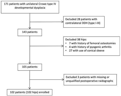
Figure 2 The measurement methods of dislocation height and distalization of greater trochanter. The dislocation height was presented as two indicators, which were perpendicular distances from the femoral head/neck junction and the tip of greater trochanter to the inter-teardrop line. The distalization of the greater trochanter was calculated by differences between the pre- and post-operative height of the greater trochanter.
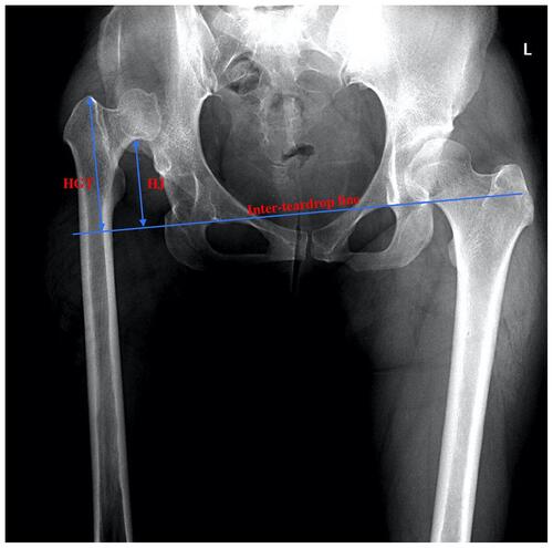
Table 1 Patient Demographics of the STO and Non-STO Groups
Figure 3 X-ray of a 42-year-old woman undergoing THA with subtrochanteric osteotomy. (A) Pre-operative X-ray. (B) Post-operative X-ray.
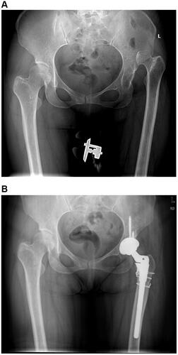
Figure 4 X-ray of a 24-year-old woman undergoing THA without subtrochanteric osteotomy. (A) Pre-operative X-ray. (B) Post-operative X-ray.
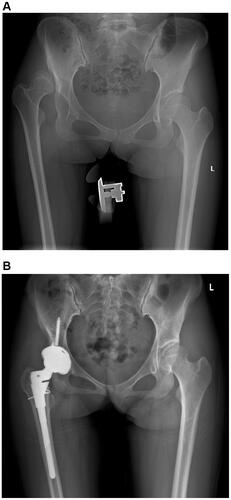
Table 2 Reliability of Length Measurements
Table 3 The Predictive Values for Three Indicators
Figure 5 ROC curves for dislocation height and distalization of greater trochanter in predicting the use of subtrochanteric osteotomy.
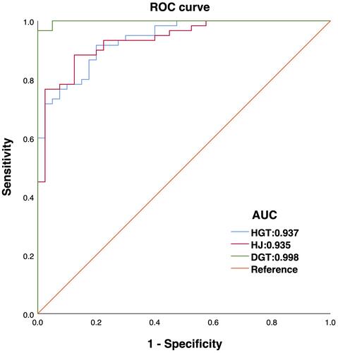
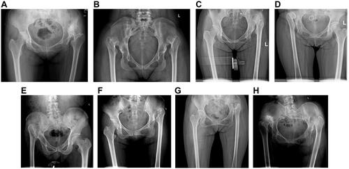
Figure 6 (A–D) The position of head/neck junction can be changeful because of the varying neck-shaft angles. (E–H) Due to the morphological complexity of femoral head, the head/neck junction is sometimes hard to measure, especially in the cases with severe abrasion or absence of the femoral head.