
Figures & data
Figure 1 An example of EP procedure. (A) Observe the papillary lesion; (B) Lift the lesion by submucosal injection (when necessary); (C and D) En bloc resection of the lesion with endoloop; (E and F) Cannulate the bile duct, and retain the guidewire in the bile duct; (G) Hemostasis with metal clips; (H) Implant the biliary stent; (I) Spray hemostatic sealant (fibrin glue) on the closed wound.
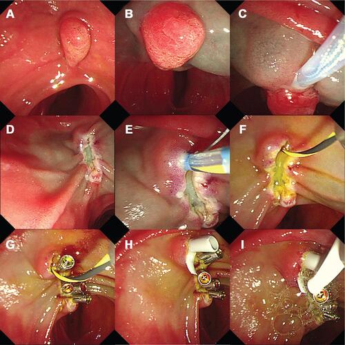
Table 1 Characteristics of Patients in the Overall Study Population
Table 2 Post-EP Complications in 76 Patients
Table 3 Univariate Analysis of Post-EP Complication Related Risk Factors
Table 4 Univariate Analysis of Post-EP Pancreatitis Related Risk Factors
Table 5 Univariate Analysis of Post-EP Bleeding Related Risk Factors
Table 6 Multivariate Analysis of Post-EP Complications