Figures & data
Table 1 Baseline characteristics
Table 2 Comparison of BCVA at different times
Table 3 Comparison of complications between the two groups
Figure 1 Images of complete healing of corneal epithelia. (A, B) Epithelial healing at 3 and 7 days after surgery in infectious central corneal ulcers; (C, D)epithelial healing at 3 and 7 days after surgery in infectious peripheral corneal ulcers.
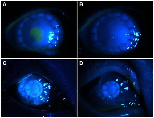
Figure 2 Cases of graft failure. (A–C) Cornea status at preoperation, rejection episode 3 months after APCS transplantation, and after treatment with PKP in infectious central corneal ulcer group. (D–F) Cornea status prior to surgery, at 3 months after APCS transplantation, and after treatment with PKP in infectious peripheral corneal ulcers. APCS, acellular porcine corneal stroma; PKP, penetrating keratoplasty.
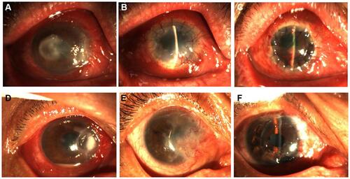
Figure 3 Representative images of graft melting. (A) At 4 months, the APCS graft had dissolved, and irreversible turbidity appeared after therapy (B) in the infectious central corneal ulcer grouper group. (C) At 3 months, the APCS graft had dissolved, and was then controlled after therapy (D) in the infectious peripheral corneal ulcer group.
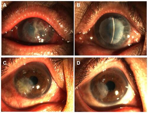
Table 4 Graft-transparency scores at postoperative 12 months
Table 5 Neovascularization scores at postoperative 12 months
Figure 4 Graft-survival rates in the two groups.
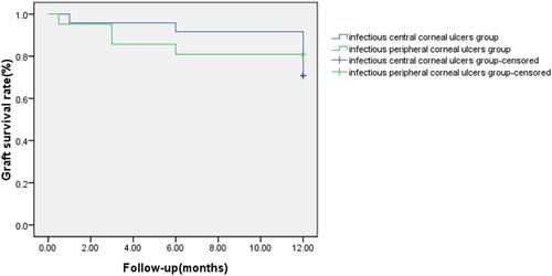
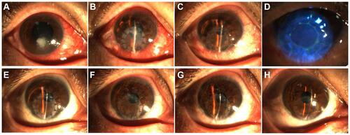
Figure 5 Successful graft transplantation in the infectious central corneal ulcer group. (A–D) Before surgery (A) and at 1 day (B), 7 days (C and, D), 1 month (E), 3 months (F), 6 months (G), and 12 months (H) postoperatively.
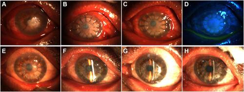
Figure 6 Another successful graft transplantation in the infectious central corneal ulcer group. (A–D) Before surgery (A) and at 1 day (B), 7 days (C and, D), 1 month (E), 3 months (F), 6 months (G), and 12 months (H) postoperatively.
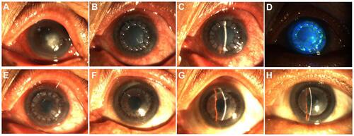
Figure 7 Successful graft transplantation in the infectious peripheral corneal ulcer group. (A–D) Before surgery (A) and at 1 day (B), 7 days (C and, D), 1 month (E), 3 months (F), 6 months (G), and 12 months (H) postoperatively.
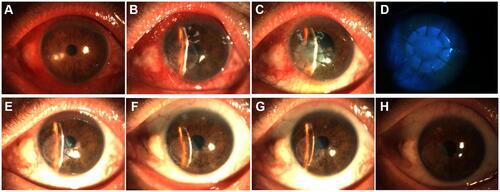
Figure 8 Another successful graft transplantation in the infectious peripheral corneal ulcer group. (A–D) Before surgery (A) and at 1 day (B), 7 days (C and, D), 1 month (E), 3 months (F), 6 months (G), and 12 months (H) postoperatively.