Figures & data
Figure 1 Defect and plate augmented with injectable bone graft substitute.
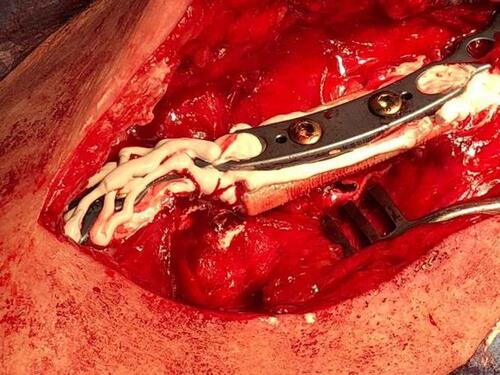
Table 1 Demographics and Baseline Characteristics
Table 2 Radiographic, Clinical and Functional Outcomes
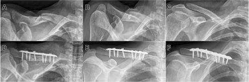
Figure 2 Displaced Allman type 1 fracture with free fragment; (A) preoperative axial radiograph; (B) postoperative axial radiograph after ORIF with a plate and screws and augmentation of both the bone defect and the plate with gentamicin-loaded bone graft substitute; (C) axial radiograph at 6 months showing bony consolidation; (D and E) coronal computed tomography (CT) scan at six months, various layers; and (F) transversal CT scan at 6 months confirming excellent bony consolidation.
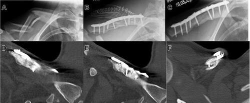
Figure 3 Refracture; (A) axial radiograph following initial surgical treatment of an Allman type 1 fracture without defect augmentation; (B) axial radiograph following implant removal; (C) axial radiograph of the refracture following implant removal; (D) post-operative axial radiograph after ORIF and defect/implant augmentation, (E) axial radiograph at 12 months revealing excellent radiographic union; and (F) axial radiograph upon implant removal at 18 months.
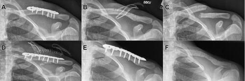
Figure 4 Delayed union after non-surgical treatment of an Allman type 1 fracture; (A) axial radiograph of a little displaced shaft fracture; (B) axial radiograph at 2 months revealing delayed union; (C) anterior-posterior (AP) radiograph confirming delayed union; (D) axial radiograph following surgical intervention with resection of the delayed union and debridement, ORIF and filling of the residual bone defect with CERAMENT G; (E) axial radiograph and (F)AP radiograph at 12 months showing excellent bony union.