Figures & data
Figure 1 Findings on imaging.
Notes: (A) Plain X-ray images show the apophysis of the bone on the lateral side of the femoral diaphysis. (B) Magnetic resonance T2-weighted fat-suppressed image shows the high signal intensity at the diaphysis. (C) Bone scintigraphy reveals an accumulation in the right femoral diaphysis.
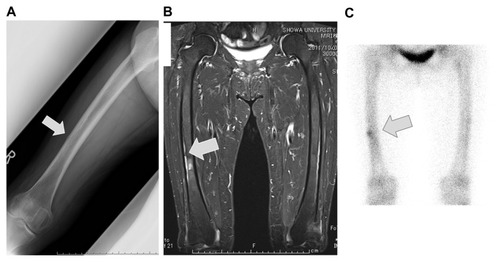
Figure 2 MMA resin-embedded V bone stain.
Notes: A slight double-labeling of tetracycline can be observed. The trabeculae were narrow, and both osteoblasts and osteoclasts were decreased.
Abbreviations: MMA, methyl methacrylate; V bone, villanueva bone.
Abbreviations: MMA, methyl methacrylate; V bone, villanueva bone.
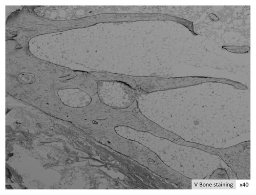
Table 1 Bone morphology measurement results