Figures & data
Figure 1 Patient presents with multiple nodules in the right auricle, a typically affected area.
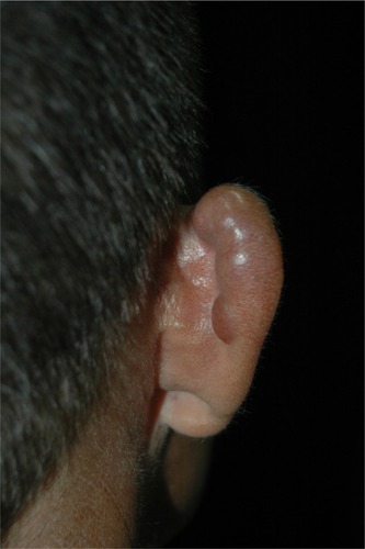
Figure 2 Typical primary fibrous appearance, with nodular plaques covered by smooth and shiny skin, with small exulcerated areas and visible telangiectasias.
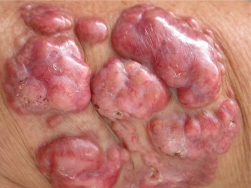
Figure 3 Local destruction of the left auricle, presenting exophytic erythematous-brownish lesion, with a pedunculated aspect and telangiectasias.
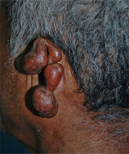
Figure 4 Superimposed carcinomatous degeneration on typical fibrous Lacaziosis nodules.
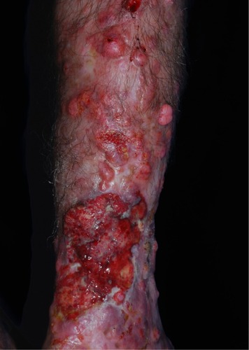
Figure 5 Ulcerated presentation with small fibrous plaques and nodules at its borders.
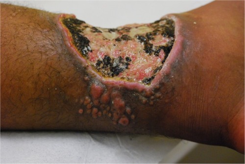
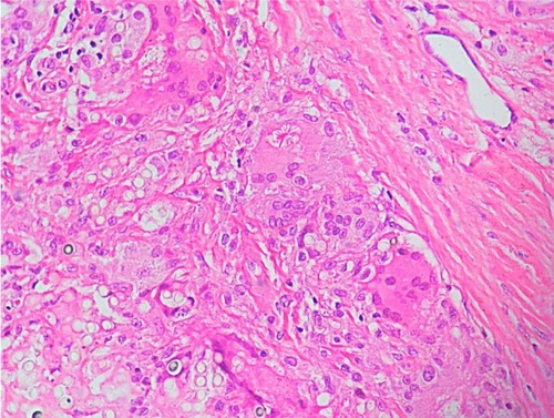
Figure 6 Histological section showing round and oval yeast-link structures with birefringent membrane, with isolated and Rosario beads distribution, commonly found in Jorge Lobo’s disease.
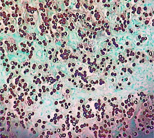
Figure 7 Histological section of a patient with lobomycosis.