Figures & data
Table 1 Baseline demographic characteristics
Figure 1 The changes of subfoveal choroidal thickness after photodynamic therapy.
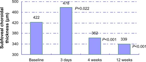
Table 2 Subfoveal choroidal thickness at baseline
Table 3 Subfoveal choroidal thickness during follow-up
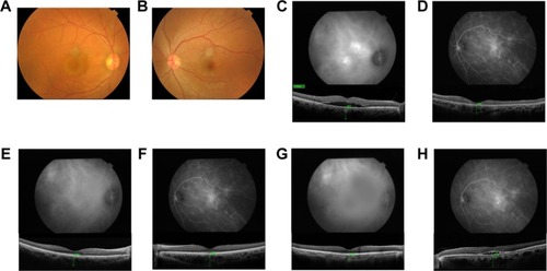
Figure 2 Case 1, a 36-year-old man with blurred vision in his right eye. (A) A fundus photograph of the right eye showing a serous retinal detachment at the fovea. (B) Fundus photograph of the left eye is normal. (C and D) ICGA showed bilateral choroidal vascular hyperpermeability in middle-phase. The subfoveal choroidal thickness at the baseline was 452 μm in the right eye and 351 μm in the left eye. (E and F) After PDT, the subfoveal choroidal thickness in his right eye decreased to 370 μm at week 4. However, the subfoveal choroidal thickness in the left eye increased to 494 μm, with ICGA indicating aggravation of his choroidal vascular hyperpermeability. (G and H) At week 11, in his left eye, ICGA showed aggravation of choroidal vascular hyperpermeability in middle-phase, with a subfoveal choroidal thickness of 470 μm.