Figures & data
Figure 1 The preoperative abdominal CT images of the cecal cancer.
Notes: (A, B, and C) Axial CT with contrast enhancement revealed cancer-affected terminal ileum, right abdominal and pelvic wall (indicated by arrows). (D) Coronal image showed a slight obstruction in the small intestine and an invasion of the abdominal wall (indicated by arrow).
Abbreviation: CT, computed tomography.
Abbreviation: CT, computed tomography.
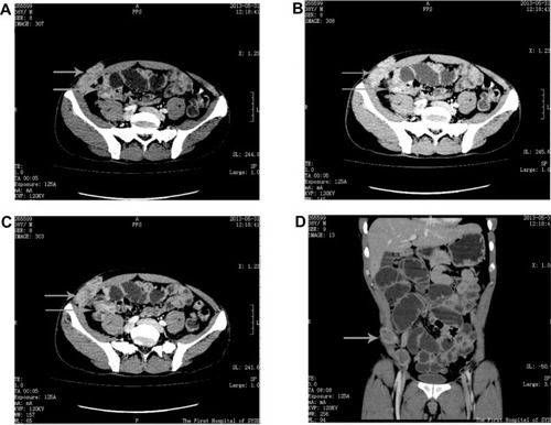
Figure 2 Intraoperative photos of the surgical process.
Notes: (A) Resected the tumor from the abdomen. (B) Resected the tumor from the abdominal wall. (C) Fixed the visceral face of the patch to the visceral peritoneum through the abdomen. (D) Fixed the fascial face of the patch to the fascia and the muscle through the abdominal wall.
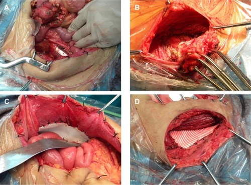
Figure 3 Photos of the right lower abdomen and specimens.
Notes: (A) The old appendectomy incision in right lower abdomen. (B) The postoperative right lower abdomen. (C and D) Pathological specimens.
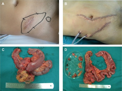
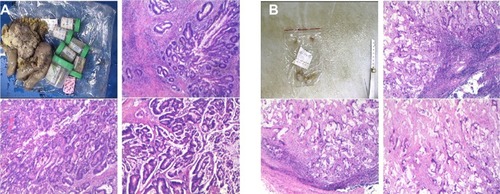
Figure 4 Photos of pathological images.
Notes: (A) The typical pathological images of the tumor. (B) The typical pathological images of the inguinal lymph nodes.