Figures & data
Figure 1 Skin lesions and histopathology.
Notes: (A) The proband and her son; (B) back of the proband; (C) the left axillary region of the proband (Crowe’s sign); (D) the inguinal region of the proband (Crowe’s sign); (E) the forehead of the son; (F) the back of the son; (G) histopathology of depigmented lesion of the proband’s right upper arm (HE ×20).
Abbreviation: HE, hematoxylin-eosin staining.
Abbreviation: HE, hematoxylin-eosin staining.
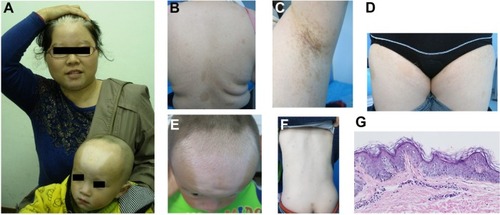
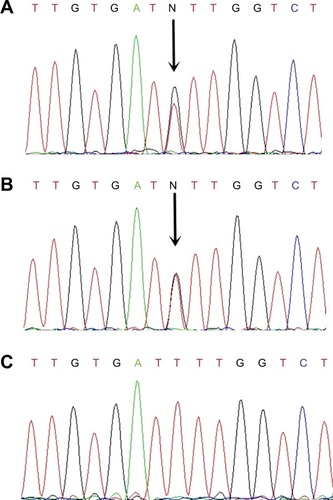
Figure 2 Mutation of the KIT gene.
Notes: (A) The proband’s KIT genomic sequence coding base numbers 2424–2438, which showed c.2431T>G heterozygous mutation; (B) the son’s KIT genomic sequence coding base numbers 2424–2438, which showed c.2431T>G heterozygous mutation; (C) the equivalent of KIT in a normal individual.