Figures & data
Figure 1 Ultrasonography image taken at 7 weeks’ gestation showed the gestational sac to be located beneath the uterine cavity, which was surrounded by rich blood flow signal.
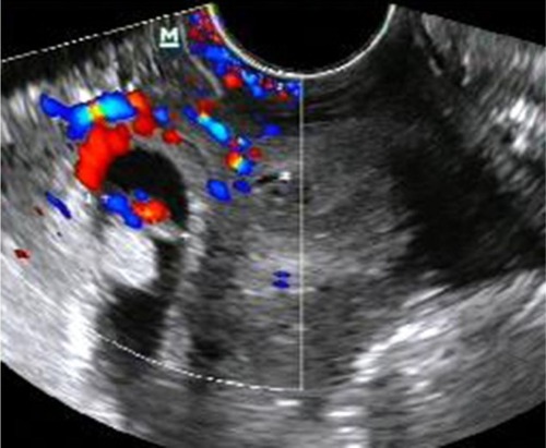
Figure 2 In laparoscopic approach, we detached bladder reflex of the uterus peritoneum.
Notes: (A) In laparoscopy, we found the violet lesion in the lower segment of uterus. (B) Partial bladder reflex of the uterus peritoneum was detached. (C) The bladder reflex of the uterus peritoneum was completely detached.
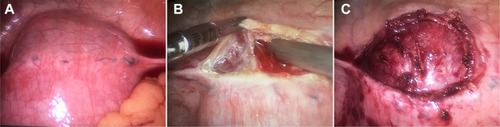
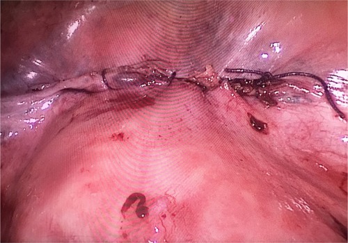
Figure 3 The uterus scar was sutured.