
Figures & data
Figure 1 (A) Port positioning. (B) A 30-day postoperative view of incision of a 38-year-old male patient.
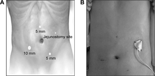
Figure 2 A 20-G IV catheter assembly. The smaller inset figure represents the 20-G needle and catheter, separated.
Abbreviations: G, gauge; IV, intravenous.
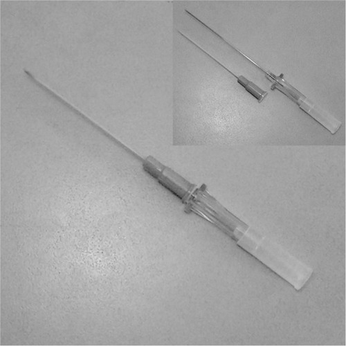
Figure 3 Demonstration of the modified laparoscopic needle catheter jejunostomy.
Notes: (A) The first anchoring stitch was placed at the 10 o’clock position of the IV catheter puncture point, taking a bite at the anterior abdominal wall and the seromuscular layer of the proximal jejunum. (B) Approximation of the bowel to the anterior abdominal wall (the IV catheter was retracted in the abdominal wall). (C) The IV catheter was advanced into the jejunal lumen. (D) Placement of the needle catheter jejunostomy into the distended jejunum. (E) A schematic view showing the purse-string suture around the feeding tube using intracorporeal suturing. (F) Intraoperative view of the placement of the purse-string suture. (G) Placement of the other two anchoring stitches around the feeding tube at the 2 and 6 o’clock position, respectively. (H) The antitorsion suture was placed approximately 5 cm distally to fix the jejunum to the anterior abdominal wall.
Abbreviation: IV, intravenous.
Abbreviation: IV, intravenous.
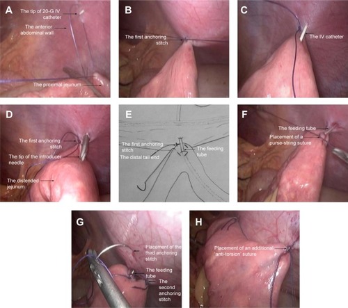
Table 1 Patient characteristics and preoperative clinical data
Table 2 Surgical details