Figures & data
Figure 1 Chest radiograph showing multiple cavitating lung lesions in a patient with Wegener’s granulomatosis.
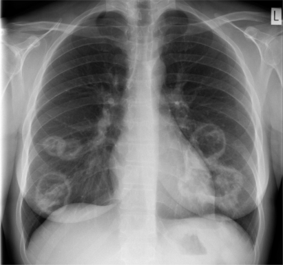
Figure 2 Renal biopsy from a patient with AAV and severe renal failure showing a glomerulus containing an extensive cellular crescent with a break in the basement membrane and surrounding fibrin deposition (arrowed) Methenamine silver stain, X400.
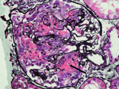
Table 1 EUVAS disease categorization of ANCA-associated vasculitisCitation9
Figure 3 Biochemical changes over time in a patient with PR3-ANCA demonstrating rapid decrease in serum creatinine, ANCA titer and C-reactive protein following initiation of immunosuppression.
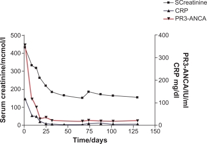
Table 2 Dose modification of pulsed intravenous CYP as used in the CYCLOPS trialCitation44
Table 3 Suggested schema for the management of ANCA-associated vasculitis, adapted and updated from Pallan et alCitation98