Figures & data
Figure 1 (A) Parasternal long axis view showing a thickened aortic wall in a patient with type A intramural aortic hematoma (arrowhead). (B) Same view with color doppler. Notice a significant aortic regurgitation.
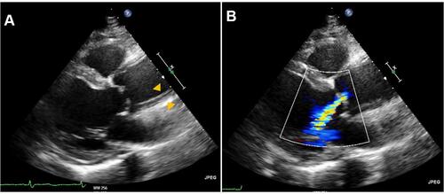
Figure 2 (A) Unenhanced computed tomography image where aortic intramural hematoma appears as a crescentic hyperattenuating thickening in the aortic wall (asterisk). (B) Same slice after contrast administration. The aortic intramural hematoma can be observed as non-enhanced crescent-shaped aortic wall thickening. Intimal calcification is characteristically displaced inward (arrowhead).
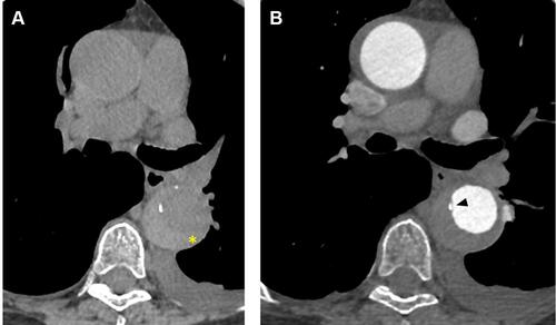
Figure 3 (A) Ulcer-like projection in the descending aorta of a patient with aortic intramural hematoma (arrow). (B) Tiny intimal disruption corresponding to an artery branch ostium (arrow).
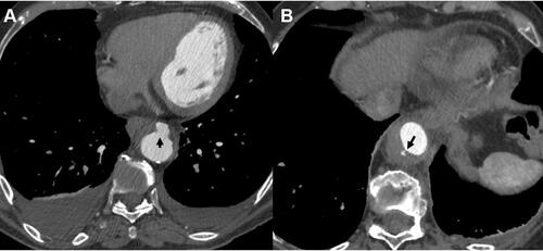
Figure 4 Two computed tomography slices showing a mural thrombus. As a difference to aortic intramural hematoma, intimal calcium remains external to the thrombus (arrow). Borders are usually irregular (arrowhead).
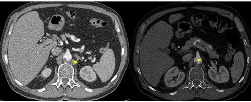
Figure 5 Incomplete dissection with some degree of sub-adventitial hematoma on a computed tomography slice. Notice the intimal tear (arrow).
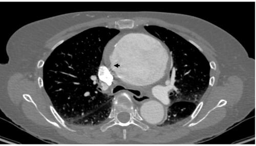
Figure 6 Magnetic resonance images (echo-gradient sequence, axial and sagittal views) showing intramural aortic hematoma. An ulcer-like projection is observed at the abdominal aorta (arrow).

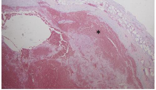
Figure 7 Histological section (hematoxylin eosin staining) of a patient with intramural hematoma. Hematoma (asterisk) within the aortic media is well documented.