Figures & data
Figure 1 Schematic drawing demonstrating various approaches to AVM. (A) Intra-operative clip or coagulation devascularization during surgical resection. (B) Endovascular nidus embolization. (C) Endovascular feeders occlusion (devascularization) prior surgical resection.
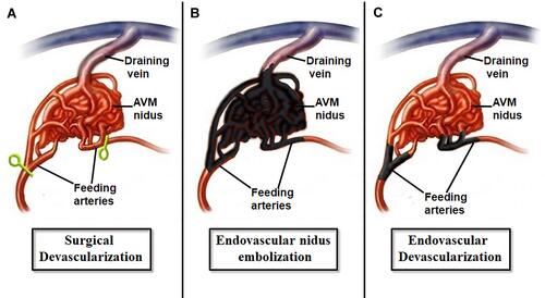
Figure 2 (A) PA left ICA, (B) lateral left ICA, (C) PA left basilar artery, and (D) lateral left basilar artery angiography showing the morphology of the splenium AVM supplied by the A5 segment of the left ACA and the lateral posterior choroidal artery branch of the left PCA and draining into the vein of Galen. (E) Lateral left ICA and (F) lateral left basilar artery angiography after occlusion of the feeding artery showing the significant regress of the AVM. (G) PA and (H) lateral skull X-rays showing the location of the Onyx® and devascularization sites.
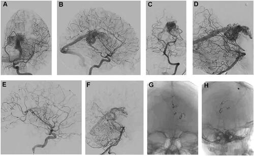
Figure 3 (A–C) Intra-operative views during surgery showing the dark embolized feeding arteries marked by Onyx® which were safely detached (arrows). Note the appearance of the nidus as a non-bleeding tuft of soft tissue. (D) Post-operative brain CT scan showing the bed of the resected AVM. Note the embolized arteries at the site of the devascularization and surgical detachment. (E–H) Post-operative cerebral angiography showing total resection of the AVM.
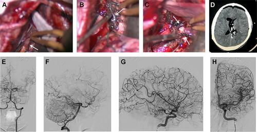
Figure 4 (A) Brain MRI showing hemorrhage in the right frontal lobe. (B and C) Cerebral angiography showing the right frontal AVM supplied by M5 branch of the right MCA and draining into a cortical vein emptying into right superficial sylvian vein. (D and E) Cerebral angiography after endovascular devascularization showing the significant regress of the AVM.
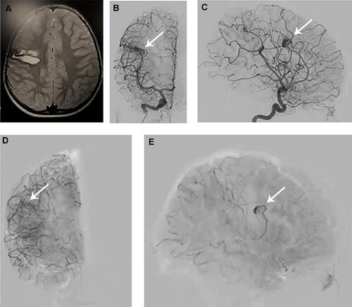
Figure 5 (A–D) Intra-operative views during surgery showing the dark embolized feeding artery which was detached (arrow). Note that the nidus was resected in piecemeal fashion. (E and F) Post-operative cerebral angiography showing total resection of the AVM.
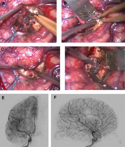
Figure 6 (A and B) Cerebral angiography of the left and right ECA’s showing the arterial supply of the scalp AVM. (C and D) Cerebral angiography of the left ICA showing the AVM supply from the supraorbital artery branch of the left ophthalmic artery. (E–G) Cerebral angiography of the bilateral ECA after endovascular devascularization of bilateral superficial temporal arteries showing the significant regress of the AVM. Note the site of the devascularization (arrows).
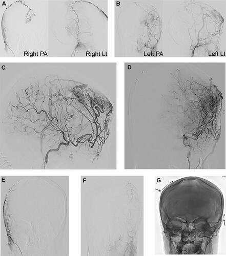
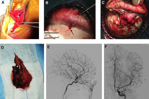
Figure 7 (A) Intraoperative picture showing the site of the left eye-brow incision and the cut of the left supraorbital artery (arrow). (B) Intraoperative picture showing the dissection of the AVM in subperiosteal plane and exposure of the draining bony venous pore (arrow). (C) Intraoperative picture showing the peri-galeal plane of the AVM dissection with noticed very minimal blood oozing from the soft tissues. (D) The AVM after resection. (E and F) Post-operative cerebral angiography of the left ICA showing the total resection of the nidus of the AVM.