Figures & data
Figure 1 (A) Preoperative radiography showing the stent position; (B) intraoperative angiography; (C) during OCT-guided atherectomy; (D) neointimal hyperplasia/plaque material was removed; (E) angiography after completion.
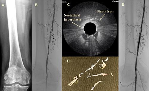
Figure 2 (A) Preoperative radiography showing the stent position; (B) intraoperative angiography; (C) during OCT-guided atherectomy; (D) neo-intimal hyperplasia/plaque material was removed; (E) angiogram after completion.
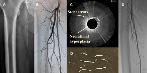
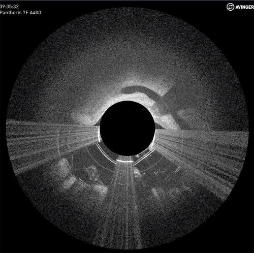
Figure 3 Local dissection during atherectomy was identified using OCT, and the flap removed. This is one of the intraoperative screens captured during case 1.