
Figures & data
Table 1 Characteristics and Clinical Presentations of Hemodialysis Patients with Central Vein Disease
Figure 1 The anatomical distribution of solitary central vein lesions in hemodialysis patients. Combined lesions (not illustrated) contributed to 24 (36.4%) of the patients as follow: 9 (13.6%) cases involved SCV + BCV, 9 (13.6%) cases BCV + SVC, 2 (3.0%) cases SCV+BCV+SVC, 1 (1.5%) case IJV + SVC, 1 (1.5%) case IJV + BCV, 1 (1.5%) case IJV + BCV + SVC, and 1 (1.5%) case SCV + IJV + BCV.
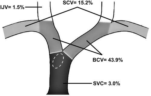
Table 2 Factors Associated with the Type of Central Vein Lesion (Stenosis Vs Occlusion) in Hemodialysis Patients
Table 3 Factors Associated with the Side of the Central Vein Lesion in Hemodialysis Patients
Table 4 Factors Associated with Installation of a Central Venous Catheter in Hemodialysis Patients with Central Vein Disease