Figures & data
Table 1 Main Available Diagnostic Methods for Evaluation of Pulmonary Valve Stenosis
Figure 1 Multimodality imaging evaluation of PS. (A) PSAX view with Color Doppler showing flow turbulence in RVOT*; (B) CW Doppler of severe PS; (C) Reduce PV opening† on SSFP cine imaging; (D) PA trunk dilatation with flow turbulence due to Jet lesion.
Abbreviations: PSAX, parasternal short axis; RVOT, right ventricle outflow tract; CW, continuous wave; PS, pulmonary stenosis; PV, pulmonary valve; SSFP, steady state free procession; PA, pulmonary artery.
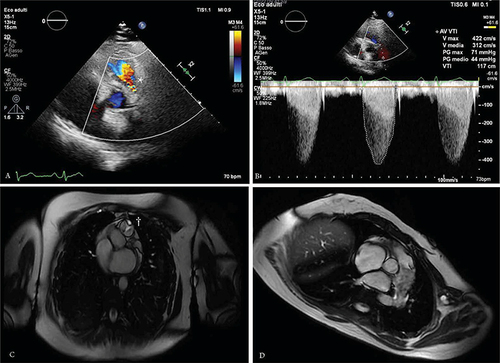
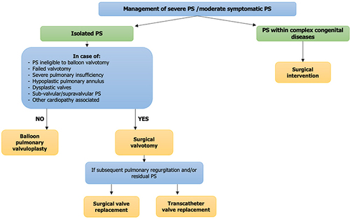
Figure 2 Flow chart for the management of patients with PS.
Abbreviation: PS, pulmonary stenosis.