
Figures & data
Figure 1 (a) Prior to treatment. (b) The plaques were lighter in color after one year of treatment.
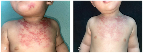
Figure 2 (a) B-ultrasound examination showed hyperechoic area with vascular echo. (b) Follow-up B-ultrasound showed scattered hyperechoic areas with a little blood flow signals.
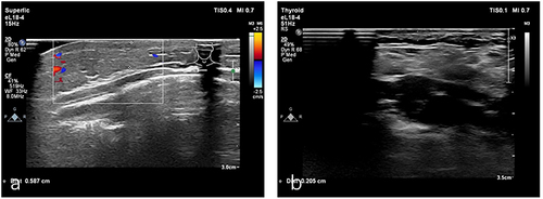
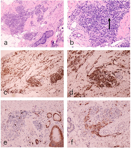
Figure 3 (a) Histopathological showing focal, well-circumscribed nodules comprised of atypical spindle cells (HE, ×50). (b) The spindle-shaped endothelial cells aligned to form a slit-like vascular channels (arrow, HE, ×100). (c) The proliferated vessels were positive for CD31(CD31, ×50). (d) The proliferated vessels were positive for CD34(CD34, ×50). (e) The proliferated vessels were negative for GLUT-1 (GLUT-1, ×50). (f) D2-40 staining was negative in the center of the lesion and positive in the surrounding dilated lymphatic vessels (D2-40, ×50).
Data Sharing Statement
The data that support the findings of this study are available on request from the corresponding author upon reasonable request.